Atopic dermatitis - diagnosis, symptoms, and treatment at EMC
Since its discovery, the disease has had more than a hundred designations, until in 1923, Coca and Cooke proposed the term "atopia" (from Greek. - strangeness, strangeness) to determine the state of hypersensitivity in hay fever, asthma, and "atopic eczema," which was later renamed "atopic dermatitis" by Wize and Sulzberger in 1933. Since then, this name has been generally accepted.
Atopic dermatitis is a multifactorial inflammatory skin disease. Its development is influenced by heredity, disorders in the immune system and an unfavorable environment.
Causes of the disease
The initial appearance of symptoms of the disease occurs under the influence of various external and internal factors, mainly in children of the first years of life. Atopic dermatitis can recur, which often leads to psychological problems in the process of personality formation and a decrease in the quality of life in adulthood.
One of the main causes of atopic dermatitis is a genetic predisposition. It has been proven that more than 20 genes can be involved in the development of allergic diseases.
According to research, atopic dermatitis develops in 80% of children whose parents suffer from this disease. More than 50% of children inherit this disease if only one parent is sick. At the same time, if atopic dermatitis is diagnosed in the mother, the risk increases 1.5 times.
Stages of disease development
Atopic dermatitis is most often manifested in childhood and has various stages of the course, the symptoms may vary depending on age.
There is no single generally accepted classification of atopic dermatitis, but there is a working version, according to which 4 stages of the disease are distinguished.
The initial stage. As a rule, it develops in children with increased vulnerability of the skin and mucous membranes, instability of water-salt metabolism, a tendency to allergic reactions and decreased resistance to various infections. The distinctive symptoms of the stage are hyperemia, swelling and peeling of the skin on the cheeks. With timely and proper therapy, it is completely cured. The symptoms do not go away on their own, with improper and untimely treatment, symptoms may worsen or the disease may enter a more severe stage.
The stage of pronounced skin changes, or the stage of progression. It almost always includes two phases: acute and chronic.
Remission stage. Disappearance or significant reduction of symptoms. The duration of the stage is from several weeks to 5-7 years. In difficult cases, the disease proceeds without remission and persists for life.
The stage of clinical recovery. According to the clinical diagnosis, the manifestations of the disease are absent for 3-7 years or more.
Forms of atopic dermatitis
Each form is characterized by the presence of itching of varying intensity.
Infant form (from birth to 2 years old). Redness and small bubbles appear on the skin, from which a bloody liquid is released when pressed. The liquid, drying out, turns into yellowish-brown crusts. The itching gets worse at night. As a result of scratching, marks and cracks appear on the skin. Symptoms most often appear on the face, may be on the arms and legs (in the elbow and popliteal folds), buttocks.
Children's uniform (3-7 years old). Redness, swelling, and crusts appear on the skin, the integrity of the skin is disrupted, the skin becomes thicker, and the skin pattern becomes more pronounced. Nodules, plaques, and erosions form. Cracks in the palms, fingers, and feet cause severe pain.
Adolescent form (8 years and older). Red plaques with blurred edges appear on the skin, pronounced dryness of the skin, and many itchy cracks. Most often, the disease is localized on the flexor surfaces of the arms and legs, wrists, backs of the feet and palms.
Prevalence of atopic dermatitis
Limited. The disease manifests itself only in the neck, wrist, elbow and popliteal folds, palms and backs of the feet. The rest of the skin remains unchanged. The itching is moderate.
Common. The disease occupies more than 5% of the body surface. The rash spreads to the extremities, chest and back. The rest of the skin takes on an earthy hue. The itching becomes more intense.
Diffuse. The entire skin surface is affected. The itching is pronounced and intense.
Severity of the current
It is estimated based on the intensity of skin rashes, the prevalence of the process, the size of the lymph nodes, etc.
Light flow. It is characterized by mild hyperemia, fluid discharge (exudation), peeling, isolated rashes, mild itching. Exacerbations occur 1-2 times a year.
The course is of moderate severity. Fluid secretion increases, and multiple lesions appear. The itching becomes more intense. Exacerbations occur 3-4 times a year.
Severe course. Multiple extensive lesions, deep cracks, and erosions appear. The itching increases and becomes permanent.
Symptoms
Clinical manifestations are characterized by erythematous, exudative and lichenoid rashes, which are accompanied by intense itching. Atopic dermatitis is often associated with abnormalities of the skin's barrier function, sensitization to allergens, and recurrent skin infections. Dysbiosis of the skin microbiota may also play a key role in the development of atopic dermatitis. It has been scientifically proven that atopic dermatitis is a skin symptom of a systemic disorder and often manifests itself as the first step in the so-called "atopic march", which causes bronchial asthma, food allergies and allergic rhinitis.
The main symptoms of atopic dermatitis
- ichthyosis, xerosis, dry skin
- palm hyperlinearity
- darkening of the skin of the eye sockets
- Hertog's sign (lack or absence of hair from the outside of the eyebrows)
- the Denier-Morgan fold (the longitudinal fold of the lower eyelid)
- persistent white dermographism
- white lichen, hair lichen
- follicular keratosis
- folds on the front of the neck
Additional symptoms:
- pallor of the face
- low hair growth limit
- delayed reaction to acetylcholine
- linear furrows on the fingertips
- keratoconus or cataract
How to diagnose atopic dermatitis?
Diagnosis of atopic dermatitis begins with a mandatory visit to a dermatologist-allergist. The extended allergodiagnostics includes laboratory blood tests for allergens, diagnosis of food intolerance, molecular diagnostics, as well as prik tests.
Molecular diagnostics is the most modern method that makes it possible to identify causally significant factors of the disease as accurately and quickly as possible, even in the most difficult cases, when other types of analyses turn out to be uninformative.
The main diagnostic criteria:
- Itchy skin
- Skin rashes: in children under 2 years of age – on the face and on the bends of the elbows and knees, in older children and adults – thickening of the skin, increased pattern, pigmentation and scratching in the area of the bends of the extremities
- Chronic course with the possibility of relapses
- The presence of atopic diseases in the family history
- The disease first appeared at the age of 2 years
Additional indicators:
- Seasonality of exacerbations (progression in autumn and winter, regression in summer)
- Exacerbation of the disease in the presence of provoking factors (allergens, food, stress, etc.)
- Increased total and specific blood IgE
- Increased levels of eosinophils (subspecies granulocytic leukocytes) in the blood
- Hyperlinearity of the palms (increased number of folds) and soles
- Follicular hyperkeratosis ("goose bumps") on the shoulders, forearms, elbows
- The appearance of itching with increased sweating
- Dry skin (xerosis)
- White dermographism
- Tendency to skin infections
- Localization of the pathological process on the hands and feet
- Eczema of the nipples
- Recurrent conjunctivitis
- Hyperpigmentation of the skin around the eyes
- Folds on the front of the neck
- The Danier-Morgan symptom Cheilitis (inflammation of the red border and mucous membrane of the lips)
To establish a diagnosis, a combination of 3 basic and at least 3 additional criteria is necessary.
Diagnosis of therapy effectiveness
Assessment of severity
- EASI: index of the area and severity of eczema (for the doctor). Assessment of prevalence in 4 separate areas (head and neck, trunk, upper extremities, lower extremities).
- POEM: eczema severity questionnaire (for patients). The patient evaluates the severity and intensity of the symptoms over the last 7 days by answering the questionnaire questions.
Assessment of the patient's quality of life
- Dermatological Quality of Life Index (DLQI). The patient evaluates the effect of the disease on the severity of symptoms, sensations, daily activity, leisure, work/academic productivity, personal relationships and treatment over a short period of time (1 week)
- The WPAI questionnaire:SHP. Assessment of the effect of atopic dermatitis on productivity during the last 7 days. How the course of the disease affected the ability to work and perform daily activities.
Complications of the disease
Atopic dermatitis is often complicated by the development of a secondary infection (bacterial, fungal, or viral). The most common infectious complication is the appearance of a secondary bacterial infection in the form of strepto– and/or staphyloderma. Pyococcal (purulent) complications manifest themselves in the form of various forms of purulent skin lesions: ostiofolliculitis, folliculitis, vulgar or streptococcal impetigo, boils.
Various fungal infections (dermatophytes, yeast-like, mold and other types of fungi) It also often complicates the course of atopic dermatitis and negatively affects the effectiveness of treatment. The presence of a fungal infection can change the symptoms of atopic dermatitis: foci with clear scalloped and raised edges appear, cheilitis often recurs, and lesions of the occipital, inguinal folds, nail bed, and genitals are possible.
Patients with atopic dermatitis are more likely to suffer from a viral infection (herpes simplex virus, human papillomavirus). Herpes infection can provoke the development of a rare and severe complication – Kaposi's herpetic eczema. Kaposi's eczema causes widespread rashes, severe itching, fever, and purulent infection. In some cases, the central nervous system and eyes are affected, and sepsis develops.
An increase in lymph nodes in the cervical, axillary, inguinal and femoral regions may be associated with an exacerbation of atopic dermatitis. This condition resolves on its own, or after adequate treatment.
Ophthalmological complications of atopic dermatitis are recurrent conjunctivitis accompanied by itching. Chronic conjunctivitis can progress to ectropion (inversion of the eyelid) and cause lacrimation.
Treatment methods
Effective treatment of atopic dermatitis is impossible without a systematic approach that includes:
- Elimination measures: prevention of contact with irritants, including in food, and household allergens.
- Regardless of the severity of the disease, if necessary, treatment is supplemented with antihistamines, antibacterial, antiviral and antimycotic agents.
- Softening and moisturizing agents to restore the impaired barrier function of the skin. It is currently known that the addition of a ceramide-dominated emollient to standard therapy leads to both clinical improvement and a reduction in water loss through the skin and an improvement in the integrity of the stratum corneum. These drugs are recommended to be used as maintenance therapy during remission.
- Topical glucocorticosteroids are the basis of anti-inflammatory treatment, showing high effectiveness in combating acute and chronic skin inflammation with limited lesions. Due to concerns about possible side effects associated with continued use, these drugs are not used for maintenance therapy.
- Calcineurin inhibitors are absolutely safe for damage to the skin of the face and eyelids. Several studies of pimecrolimus cream have revealed that the use of the drug at the earliest stages of the disease leads to a significant reduction in the need for "rescue" therapy with glucocorticosteroids.
- In moderate cases, the use of phototherapy is important. It affects inflammatory cells (neutrophils, eosinophils, macrophages, Langerhans cells) and alters cytokine production, and also has a persistent antibacterial effect. Moreover, phototherapy with UV rays can have a normalizing effect on the immune status.
- In severe atopic dermatitis, in addition to topical remedies, treatment includes the use of systemic short-course glucocorticosteroids and cyclosporine. In 55% of cases, a positive effect occurs after 6-8 weeks of use. Continuous therapy is not recommended for more than 1-2 years because cyclosporine has potential toxicity. Biological therapy in the treatment of atopic dermatitis.
Dupilumab is the world's first monoclonal antibody-based treatment for atopic dermatitis. This is a class of drugs that have high selectivity for key components of the pathological process. Antibodies have the ability to precisely bind to an antigen due to special antigen-binding sites that have high specificity for it. For antibody-based drugs, this determines their selectivity for a specific target. New drugs work where previous drugs are powerless. For this reason, they can be more effective or used in cases where the disease has proven to be resistant to traditional drugs.
It has also recently been revealed that omalizumab is the most effective drug in the treatment of allergic asthma and allergic rhinitis. Thus, it can potentially neutralize the effect of immunoglobulin in atopic dermatitis.
Prevention of atopic dermatitis
With atopic dermatitis, even quite harmless factors, such as clothing and moisture, can provoke itching. That is why it is recommended to wear natural fabrics and avoid intense physical exertion. Washing powders, even in small quantities remaining on linen and clothes, can irritate the skin, therefore it is recommended to use hypoallergenic soap-based washing powders, adding a repeated cycle of rinsing the laundry. You should also exclude products containing perfumes and preservatives, and try new cosmetic products on a small area of the skin before use.
It is necessary to cleanse and moisturize the skin, because with atopic dermatitis, due to disorders in the lipid metabolism of the skin, its dryness increases. Violation of the protective function can cause the development of secondary bacterial, viral and fungal infections. That is why the skin with atopic dermatitis requires special care: shower cleansers ("soap without soap"), as well as bath oils; after washing, without rubbing the skin, you must immediately apply a moisturizer. Moisturizers and balms can be applied several times a day.
Primary prevention
Primary prevention is aimed at preventing the development of atopic dermatitis and should be carried out during pregnancy. It is known that the disease is inherited, and if both parents have it, the probability of an unborn child getting sick is 60-80%.
It is recommended to exclude highly allergenic foods such as chocolate, citrus fruits, honey, nuts, etc. from the diet of a pregnant woman. On the other hand, the diet should be varied, and one-sided carbohydrate nutrition should be avoided. It is important to treat gestosis in a timely manner, which significantly increases the permeability of the placenta-fetus barrier and promotes allergization. It is recommended to limit the drug load, as many medications can cause allergies. Elimination of overloads at work and occupational hazards during pregnancy.
Equally important is prevention after the birth of a child, where breastfeeding plays a very important role, since breast milk is maximally adapted to the needs of the newborn and does not contain foreign proteins to which allergies may occur.
A child with a predisposition to atopic dermatitis should receive medication only for well-founded indications, since medications can act as allergens that stimulate the release of immunoglobulin.
Secondary prevention
It is performed when a child is diagnosed with atopic dermatitis and is designed to reduce the number of exacerbations and improve the quality of life.
Exacerbations of atopic dermatitis can be caused by:
- house dust mites;
- mold that forms on the soil of domestic plants and in damp rooms;
- components of cosmetics or detergents;
- animal hair, etc.
In this case, regular wet cleaning (including using vacuum cleaners), frequent change of bed linen, treatment of tiled walls with antifungal solutions, and the use of clothing made from natural fabrics (except wool) will help.
You can make an appointment with an EMC dermatologist by phone: +7 (495) 933 66 55.
Why the EMC
The first and only clinic in Russia, created in the image of the world's leading clinics
EMC is a multidisciplinary center offering patients a high level of medical services and a personalized approach
Worldwide recognition and awards
 Learn more
Worldwide recognition and awards
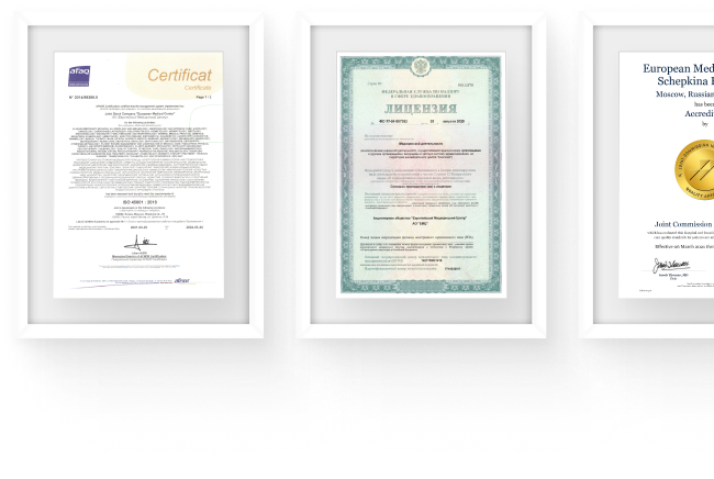
 Certificates and licenses
Make an appointment for a consultation
Specify your contacts and we will contact you to clarify the details
Reviews
and new products of the EMC


.webp)
.webp)
.webp)


