Service, direction or doctor...
Popular queries
Close search
EN
RU

Duchenne muscular dystrophy

About pathology
Causes of occurrence
Stages of the disease
Diagnostics
Treatment of Duchenne dystrophy

About pathology

Duchenne Muscular Dystrophy (DMD)It is a progressive hereditary disease that leads to weakness and muscle atrophy.

Causes, symptoms, and treatment options. They are a hereditary group of progressive myopathic disorders that occur due to defects in a number of genes necessary for normal muscle function. The main symptom is muscle weakness [6].

Distinctive features of the disease

Duchenne muscular dystrophy is one of the most severe forms of hereditary myodystrophy. The pathology is caused by a mutation in the DMD gene, which programs the protein dystrophin. In 60% of cases, the disease is inherited by boys from female carriers.

Duchenne muscular dystrophy belongs to orphan diseases, that is, it is very rare, it occurs in about one out of 3500 to 5050 newborn boys. Nevertheless, among other rare diseases, the incidence of DMD is quite high — 20% of all cases of orphan diseases occur specifically in it [1,7].

Dystrophinopathies are inherited as X-linked recessive traits and have various clinical features. For the purposes of diagnosis and treatment, dystrophinopathies are usually divided into the following categories:

  • Duchenne muscular dystrophy (DMD)

    It is associated with the most severe clinical symptoms.

  • Becker muscular Dystrophy (MDD)

    It has similar manifestations to DMD, but usually a later onset and milder clinical course.

  • Mild MDD or severe MDB

    Patients with an intermediate phenotype can be clinically classified as having either a mild form of MDD or a severe form of MDB.

  • Dilated cardiomyopathy (DCM) associated with DMD

    This is a term used when the heart is primarily affected, while the skeletal muscles remain intact.[6]

The difference between DMD and MDB is that in Duchenne disease, the protein dystrophin is not synthesized at all, while in Becker's myodystrophy, a small fragment of the protein is still formed, which is associated with a milder course.

Causes of occurrence

Dystrophin is one of the largest genes in the human genome, accounting for 0.1% of the total human genome. The DMD dystrophin gene is the largest gene identified in humans. The protein product is also extremely large, weighing 427 kilodaltons (kD). Due to the large size and complexity of the gene, it becomes a major target for mutations to date, over 4,700 of their types have been identified [2]. In turn, mutations in the b gene significantly disrupt cell function, and over time, pathological processes steadily progress, exacerbating each other.

The functions of the dystrophin gene include:

  • supports the integrity of the muscle membrane (binds the actin skeleton to the extracellular matrix);

  • support for the proper functioning of proteins during their positioning in the complex.

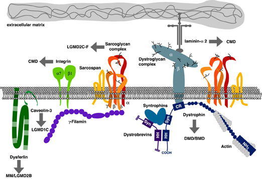
Protein complex associated with dystrophin

Rice.1. A protein complex associated with dystrophin.

Dystrophin is located on the cytoplasmic side of the plasma membrane of muscle fibers. The arrows point to the protein components that have mutated in various muscular dystrophies. Changes in the dystrophin gene cause Becker and Duchenne muscular dystrophies. Dystrophin usually provides mechanical reinforcement of the sarcolemma and stabilizes the glycoprotein complex, thereby protecting it from degradation. In its absence, the glycoprotein complex is digested by proteases. The loss of these membrane proteins can initiate degeneration of muscle fibers, which leads to muscle weakness [8].

In patients with Duchenne muscular dystrophy, the presence of dystrophin is not detected by any DNA diagnostic methods. The loss of such an important element of the chain leads to serious consequences:
  • a cascade of inflammatory reactions is triggered;
  • the cytoskeleton collapses;
  • the membrane of muscle cells loses stability and is easily damaged;
  • the ability of muscle vessels to dilate is impaired, which leads to their ischemic damage;
  • neuromuscular synapses are disrupted;
  • muscle cells lose their ability to regenerate and die.

Clinically, these changes manifest themselves as increasing muscle weakness and gradual replacement of connective and fatty muscle tissue, which eventually leads to patient disability and early death.

Stages of the disease

The manifestation of DMD symptoms usually occurs at the age of 3 to 4 years. In families where the disease occurs for the first time, the average age of pathology diagnosis is 4 years and 10 months [3]. If the patient's condition is monitored more closely due to a burdened medical history, specialized DNA tests help to identify the pathology at an earlier stage.

In early childhood, boys usually do not differ in the level of motor development from their healthy peers, or the deviations are small and are regarded as a variant of the norm. In some cases, children later begin to sit and stand up, are more clumsy at the beginning of walking, and often stumble.

The pathology begins to manifest itself more vividly after 2 years and starts with the muscles of the hip complex and neck flexors. The following stages of the disease are distinguished:

  • Preclinical stage

    There are no characteristic symptoms of dystrophy. The alertness of parents and doctors can cause deviations in the development of speech and movements (for example, abnormal gait, difficulty climbing stairs).

  • Early outpatient stage

    The patient retains the ability to move independently, but signs of muscle weakness become noticeable. It is difficult for a boy to jump or climb stairs, he often falls, walks on tiptoes or spreads his legs wide ("duck walk"). A characteristic feature is considered to be the Govers technique — in order to get up from a prone position on the floor and fully straighten up, the boy is forced to use his hands on the floor and on his own knees. There is usually a visual increase in the calf muscles (the so-called pseudohypertrophy) associated with the replacement of the lower leg muscle tissue with more voluminous connective and fatty tissue.

  • Late outpatient stage

    As the muscle fibers break down, the symptoms increase. The boy's ability to walk on stairs decreases, and he uses his hands to get up from a sitting position. Problems with posture are increasing — scoliosis, lumbar lordosis (a deep forward bend of the spine in the lower back) occurs. There is a decrease in the functionality of deep tendon reflexes (sometimes they are lost by the age of 6). During periods of forced immobility, for example, due to increasing illnesses or surgical operations, muscle weakness progresses at an accelerated rate.

  • Early outpatient stage

    The child retains the ability to move independently only for short distances (up to 10 m), but is forced to use a wheelchair. Skeletal disorders are formed: "wing-shaped" shoulder blades separated from the back, curvature of the spine, curvature of the chest. The motor activity of the hands usually persists until a late stage, that is, the child is able to independently control a wheelchair. At this stage, the likelihood of dangerous secondary disorders of the respiratory and cardiovascular systems increases.

  • Late non-ambulatory stage

    The patient loses the ability to control his hands, maintain body posture, and needs respiratory support.

Diagnostics

Due to the rarity of the disease, most doctors in conventional medical centers in their professional activities never encounter Duchenne disease, which makes it difficult to detect it. Muscle weakness as a symptom can remain unrecognized for a long time, as parents complain of impaired coordination, fatigue, delayed intellectual or linguistic development of children, non-specific symptoms that may accompany other, more common diseases. Because of this, the opportunity for early diagnosis and timely access to specialized specialists is missed.

Diagnostic methods

Physical examination

The neurologist conducts a survey of the patient and his parents, paying special attention to characteristic complaints that may indicate muscle weakness: increased fatigue, frequent falls or clumsiness, muscle and back pain, delayed intellectual and linguistic development, learning difficulties, etc. The doctor evaluates the patient's gait, examines muscle tone, strength, tendon reflexes, conducts the Govers test (reveals auxiliary techniques when lifting from the floor). The condition of the osteoarticular system and the presence of spinal curvature are assessed.
Laboratory tests

The main early sign of dystrophy is an increase in creatine phosphokinase (CK) in the blood. It is an enzyme found in skeletal muscles and is an indicator of their breakdown. With muscular dystrophy, the level of CFRP can be increased tens or hundreds of times relative to the norm. At the same time, changes in blood biochemistry appear even before any symptoms appear, due to which Duchenne muscular dystrophy is sometimes detected accidentally during a routine examination. However, as the amount of muscle tissue decreases, the level of CK decreases. At the same time as CK, increased levels of AST, ALT, and LDH enzymes may indicate possible muscular dystrophy, but this sign is also characteristic only of children under 3 years of age.

Genetic analysis

DNA testing can detect abnormalities in the dystrophin gene. Large deletions and duplications in the DMD gene are being identified and the gene is being sequenced to detect small and point mutations. This method shows up to 98% of genetic mutations.

Muscle biopsy

Immunohistochemical examination of the tissue of the gastrocnemius muscle or quadriceps femoris for the presence of dystrophin, signs of necrosis of muscle fibers and replacement of muscle tissue with fat and connective tissue. Usually, a biopsy is performed only in the absence of a definitive diagnosis based on genetic analysis.

Instrumental examinations

In young children, noninvasive examinations can be performed instead of biopsies — MRI or ultrasound of muscle tissue, wave elastography.

Additional examinations

To detect concomitant diseases, ECG, EchoCG, spirometry, radiography are prescribed, and visits to a cardiologist, endocrinologist, pulmonologist, and other specialized specialists are recommended.

Treatment of Duchenne dystrophy

The goal of treatment for Duchenne muscular dystrophy is to slow down the progression of the disease, reduce the risk of concomitant pathologies, and prolong the patient's ability to move independently. Significant progress has been achieved in this regard: whereas 30 years ago children were confined to a wheelchair until the age of 6-10, today some patients retain the ability to walk after the age of 14.

The main methods of treatment [1,4]:

  • Glucocorticoids

    They are indicated for children with DMD and should be initiated before significant physical deterioration occurs [9]. Hormonal treatment has an anti-inflammatory effect and is the basis of pharmacological treatment of DMD due to its effect on improving motor and lung function, reducing the risk of scoliosis, slowing the loss of the ability to move and possibly slowing the progression of cardiomyopathy and improving survival.

  • Cardioprotective therapy

    Drugs for the prevention of cardiovascular diseases. DMD is often accompanied by cardiomyopathy, which can reach severe stages and lead to death. The patient's cardiovascular system is regularly monitored, and if necessary, cardiopreparations are prescribed: ACE inhibitors and antiarrhythmic therapy, etc.

  • Cardiometabolic therapy

    Prescribed at the stage of decompensation, the doctor may recommend taking cardiotonics such as L-carnitine, coenzyme Q10.

  • Physical therapy and physical therapy

    They help slow down muscle degradation and prolong patients' motor activity. Patients also need speech therapy sessions in accordance with their individual needs and the stage of the disease.

  • Metabolic therapy

    Vitamin and mineral supplements (D, B, calcium) are prescribed to improve metabolic processes and prevent osteoporosis.

  • Gene therapy

    The world is actively developing drugs that can help stop a gene mutation that blocks the synthesis of dystrophin. In particular, the drug Ataluren has been developed in the USA for this purpose (Translarna, PTC Therapeutics, USA). It is able to bind to ribosomal RNA, inhibits the development of mutations, and allows for the restoration of dystrophin synthesis and translation even in the presence of already damaged genes. Other developments are also underway. So, scientists and doctors are trying to create a universal drug or method that will help with any type of gene mutation. Such a drug can be called mini and microdystrophin or the CRISPR/Cas9 method [1].

The EMC Clinic uses a progressive multidisciplinary approach to manage patients with myodystrophy. Our doctors have completed internships in the best Russian and foreign clinics, and have experience using the latest drugs and medical treatment protocols.

You can make an appointment for a consultation or ask any questions about our services online or by phone +7 495 933-66-55.

Sources

  1. Charitable Foundation for Children with Duchenne myodystrophy and Other severe neuromuscular diseases mymiofond.ru
  2. Gainetdinova Dina Damirovna, Novoselova Anastasia Andreevna MODERN POSSIBILITIES OF DIAGNOSIS AND TREATMENT OF DUCHENNE MUSCULAR DYSTROPHY // Kazan Medical University. 2020. #8470; 4. cyberleninka.ru
  3. Clinical recommendations. Progressive Duchenne muscular dystrophy. Becker's progressive muscular dystrophy www.pediatr-russia.ru
  4. Parent project "Muscular Dystrophy" (USA) www.parentprojectmd.org
  5. GremyakovaT.A., ArtemyevaS.B., BaibarinaE.N., VashakmadzeN.D., GuzevaV.I., GusakovaE.V., Kuzenkova,L.M., Lavrova,A.E., Lvova,O.A., Mikhailova, S.V., Nazarenko, L.P., Nikitin, S.S., Polyakov A. V., Dadali E. L., Rumyantsev A. G., Sakbayeva G. E., Suslov V. M., Gremyakova O.I., StepanovA.A., Shakhovskaya N.I. Consensus on the concept of modern effective therapy of Duchenne muscular dystrophy // Neuromuscular diseases. 2023. № 2. cyberleninka.ru
  6. www.uptodate.com
  7. Romitti PA, Zhu Y, Puzhankara S, James KA, Nabukera SK, Zamba GK, Ciafaloni E, Cunniff C, Druschel CM, Mathews KD, Matthews DJ, Meaney FJ, Andrews JG, Conway KM, Fox DJ, Street N, Adams MM, Bolen J, MD STARnet. Prevalence of Duchenne and Becker muscular dystrophies in the United States. Pediatrics. 2015;135(3):513. Epub 2015 Feb 16.
  8. Ervasti JM, Ohlendieck K, Kahl SD, Gaver MG, Campbell KP. Deficiency of a glycoprotein component of the dystrophin complex in dystrophic muscle. Nature. 1990;345(6273):315.
  9. Birnkrant DJ, Bushby K, Bann CM, Apkon SD, Blackwell A, Brumbaugh D, Case LE, Clemens PR, Hadjiyannakis S, Pandya S, Street N, Tomezsko J, Wagner KR, Ward LM, Weber DR, DMD Care Considerations Working Group. Diagnosis and management of Duchenne muscular dystrophy, part 1: diagnosis, and neuromuscular, rehabilitation, endocrine, and gastrointestinal and nutritional management. Lancet Neurol. 2018;17(3):251. Epub 2018 Feb 3.

Make an appointment for a consultation and we will contact you to clarify the details.

Select a call time
I have read and agree with the Personal Data Processing Policy

Why the EMS

The first and only clinic in Russia, created in the image of the world's leading clinics

EMC is a multidisciplinary center offering patients a high level of medical services and a personalized approach

600
world-renowned doctors
57
treatment directions
35
years taking care of your health
24/7
we work at any convenient time

World recognition and awards

Our achievements are confirmed by prestigious international awards
More details

World recognition and awards

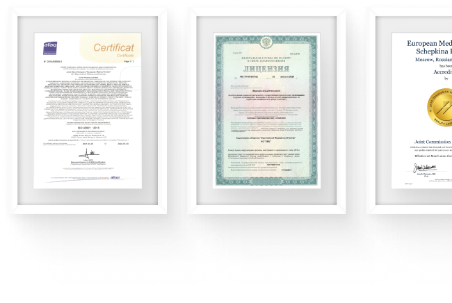
We work according to international standards, we have licenses and certificates
Certificates and licenses

Make an appointment for a consultation

Specify your contacts and we will contact you to clarify the details.

Select a call time
I have read and agree with the Personal Data Processing Policy

Questions and answers

How many people live with Duchenne muscular dystrophy?
The results of early studies on the treatment of DMD were disappointing – patients died in adolescence from respiratory tract infections and cardiomyopathy. According to modern research, with high-quality care provided by multidisciplinary teams of doctors and symptomatic treatment, with the development of
technologies for the care of respiratory and cardiac function, the average life expectancy has increased to 35 years; in some cases, patients with DMD live for more than 50 years [5].
...ещё
What diseases are possible with Duchenne dystrophy?
Autism spectrum disorders and epilepsy are more often diagnosed with myodystrophy than the general population. Already in adolescence, cardiomyopathies can manifest themselves — heart pathologies that are often accompanied by dangerous arrhythmias or sudden death syndrome. Other cardiopathologies are also possible:
persistent tachycardia, heart failure, atrial and intra-atrial conduction disorders. Due to the peculiarities of the course of myodystrophy, patients are more susceptible to infectious diseases (ARVI, pneumonia), which can pose a significant danger to them. Even before the loss of the ability to move independently, patients often have osteoporosis, complications from the gastrointestinal tract.
...ещё
Is it possible to cure muscle dystrophy?
Currently, treatment methods are being actively developed that will restore dystrophin expression, clinical studies of chemicals are underway to slow down or interrupt the pathological processes inherent in DMD, and the search for effective gene editing methods continues.

Subscribe to the newsletter
Find out before others about the special offers
and new products of the EMC
Email
Direction