Fracture of the eye socket wall
Tells Roman Kartashov,
maxillofacial surgeon
Injuries to the skull can be accompanied by fractures of the bones forming the orbit, the cavity in which the eyeball is located. If you or your child have suffered an injury that has caused a "bruise" under the eye, it is important to contact a specialist in time to determine if there is a fracture. This is important because a fracture of the orbital wall can lead to unpleasant consequences associated with impaired visual function.
Symptoms of fracture of the orbital wall
- the appearance of pain, swelling, or bruising in the eye area;
- numbness of cheeks, gums;
- nosebleed;
- eye position change, double vision;
- decreased visual acuity;
- disfiguration of the face (due to fractures of the facial skeleton with extensive injuries).
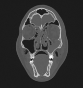
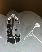
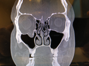
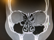
Diagnosis of fracture of the orbital wall
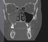
Computed tomography (CT) is the gold standard in the diagnosis of orbital fractures, allowing accurate reproduction of the skeleton of the orbit and adjacent structures in several planes. 3D reconstruction provides reliable information about the number of bone fragments of the orbit and their position.
Treatment of fractures of the orbital wall
- antibacteriale to prevent the development of infection;
- symptomatic for relief of pain, edema, subcutaneous hematomas;
- surgical to restore the former shape of the skeleton, the position of the eye, drainage of intraocular hematomas.
Fractures of the orbit differ significantly both in location and severity. Globally, it is important for a doctor to determine whether surgery is necessary.
Indications for surgery:
- visual impairment (often due to a fracture, the eye is displaced, resulting in double vision; an increasing hematoma can lead to optic nerve atrophy and vision loss; bone fragments can interfere with muscle contraction, which leads to limited eye movement);
- a violation of the structure of the facial skeleton due to displacement of bone fragments, which is manifested by changes in facial features, asymmetry, and disfigurement; compression of the suborbital nerve by dislocated bone fragments, which led to numbness of the cheeks, half of the nose, lips and gums.
It is preferable to perform surgery immediately after the fracture before the edema develops. If the swelling does appear, then it is necessary to wait 3-5 days.
If a fracture is left untreated, then adverse consequences in the form of post-traumatic deformity, visual impairment, and changes in the position of the eye are possible. They are much more difficult to treat than a "fresh" injury.
Types of operations for fractured eye sockets:
The walls of the eye socket are represented by very thin bones, and it is not possible to return them to their previous position. However, surgeons have several materials for prosthetics in their arsenal: the patient's own bone from the cranial vault, titanium mesh and various synthetic prostheses.
If we are talking about a fracture of the edge of the eye socket, then the restoration of its shape is performed by fixing titanium plates with screws, because in this place the bone is thick enough.
Rehabilitation after fracture of the orbital wall
After the operation, a CT scan is performed to monitor the result of the operation. The patient is under the supervision of a maxillofacial surgeon and an ophthalmologist.
As a rule, with a smooth course of the postoperative period, on 3-4 days the swelling in the area of surgery begins to subside, and after 7-10 days only traces of hematomas may remain.
Rehabilitation is aimed at restoring the function of vision. Patients are advised to perform oculomotor exercises, avoid increasing pressure in the nasal cavity during sneezing and blowing their nose.
Contraindications to surgery
- severe traumatic brain injury
- concomitant pathology, in which any surgery is contraindicated.
Why the EMC
The first and only clinic in Russia, created in the image of the world's leading clinics


.webp)


.webp)

Make an appointment for a consultation
Specify your contacts and we will contact you to clarify the details

and new products of the EMC