Pyoderma - what is it?
Every person is confronted with pustular skin lesions throughout their life. Impetigo, acne, sycosis, boils, diaper rash, erysipelas are an incomplete list of forms of pyoderma, which often cause a lot of inconvenience. Some types are easily transmitted by household contact.
Pyoderma is caused mainly by staphylococci and streptococci, turning the normal microflora of the skin into pathogenic.
In most cases, staphylococci infect hair follicles, sebaceous and sweat glands, causing a purulent-exudative inflammatory reaction followed by the formation of a pustule. Streptococci infect the skin without penetrating into its appendages, and cause a serous-exudative inflammatory reaction with the formation of a flare. An important role in the development of pyoderma is played by the condition of the skin and the general condition of the body.
Pyoderma can be primary or secondary. The first develops as an independent disease, the second as a complication of other skin diseases.
They increase the risk of developing pyoderma:
-
dry skin;
-
cracks;
-
microtrauma and scratching (for example, with dermatoses);
-
active exposure to chemicals (including detergents);
-
non-compliance with personal hygiene rules.
The following can provoke the development of pyoderma: immunodeficiency states, metabolic disorders, deficiency of vitamins and minerals in the body.
Pyoderma is most dangerous in newborns, as infection can generalize and sepsis can develop. The protective mechanisms in children of the first year of life cannot yet give a full-fledged immune response. Skin features are also of great importance: newborns have a thin and loose stratum corneum, the direct location of the ducts of the sweat glands and the alkaline environment of the skin surface. All this also contributes to the spread of the disease. Additional factors that may affect the development of pyoderma in newborns are overheating, rare diaper changes, and hygiene violations.
In preschool and school-age children, the same complications are possible after streptoderma as after other streptococcal infections (sore throat, scarlet fever), and the development of glomerulonephritis is also possible.
Important! Treatment of pyoderma should be carried out under the supervision of a doctor, self-medication can cause an exacerbation of the disease and subsequent complications, therefore it is strictly contraindicated.
Types and characteristics of pyoderma
Superficial pyoderma:
I. Staphyloderma
II. Streptoderma
-
Streptococcal impetigo:
streptococcal infection;
interriginal;
bullous;
ring-shaped;
syphilis-like;
surface panel.
-
Dry streptoderma
III. Strepto–staphyloderma
Deep pyoderma
I. Staphyloderma
Deep folliculitis
Furuncle, furunculosis
Carbuncle
Hydradenite
II. Streptoderma
-
Cellulite:
Ectima vulgaris
III. Strepto–staphyloderma
Clinical manifestations
Staphyloderma
Folliculitis is a purulent inflammation of the hair follicle.
Ostiofolliculitis is a small follicular superficially located cone-shaped pustules with purulent heads. Localization: the area of sebaceous hair follicles. The edges of the pustule are framed by a pink rim, 1 mm wide. More often, ostiofolliculitis occurs on the face, trunk, and limbs. After 3-5 days, crusts form in place of the pustules, which then disappear.
Superficial folliculitis is characterized by the large size and depth of the lesion of the hair follicle. The redness zone around the pustule is 2 mm. The pustule is dense, the pus is thick, yellowish-green in color. Soreness in the rash areas disappears after opening the pustules.
Deep folliculitis – painful pustules up to 1.5 cm in diameter, completely engulfing the hair follicle. The difference from a boil is the absence of a necrotic rod. With a large area of skin damage, a deterioration in the general condition may occur: fever, headaches, abnormalities in laboratory blood parameters.
Sycosis vulgaris is a chronic purulent process characterized by inflammation of the hair follicles in the nasolabial region, neck and chin, less often in other areas. It occurs more often in men. The development of sycosis can be influenced by frequent shaving, chronic rhinitis, disorders of the nervous system, internal organs and endocrine glands, hypersensitivity to staphylococci and their waste products, the presence of foci of infection (caries, chronic tonsillitis). The disease is characterized by multiple rashes of painful, sometimes itchy ostiofolliculitis and folliculitis containing pus. The foci merge, forming large areas of inflammation. The process is characterized by a chronic course. The general state of the patient's health does not change, but aesthetic defects can cause significant discomfort.
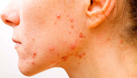
Acne vulgaris is a chronic disease affecting the sebaceous glands. They usually appear during puberty and are associated with a genetic predisposition, decreased immunity, endocrine disorders, focal infection, and diseases of the gastrointestinal tract. Excessive consumption of carbohydrates also affects the development of the disease.
The most common localization is the skin of the face, chest and back, with severe course of the process involving the shoulders and upper third of the forearms. As a result of blockage of the mouths of the hair follicles, comedones (black dots) form, then a papular form develops, manifested by pink-red hemispherical or conical papules. As a result of the addition of staphylococcal infection, a small pustule forms in the center, which dries out to form a crust or opens (pustular form). A complication of the disease is necrotic acne, when necrosis develops deep in the follicle, followed by the formation of a scab and a smallpox-shaped scar. These types of rashes are more common on the forehead and temples. A special form of the disease is conglobate (spherical) acne. They occur mainly in men and are localized mainly on the back. They are characterized by the formation of large, often multiple comedones surrounded by an inflammatory infiltrate, with the formation of a flaccid abscess, the formation of a fistula and long-term non-healing ulcerative surfaces.
Furuncle, furunculosis is a deep staphyloderma characterized by purulent-necrotic inflammation of the hair follicle and surrounding tissues. The main localization is on the face, neck, shoulders, hips or buttocks. At the beginning, redness and swelling appear in the area of the hair sac. Then the lesion turns into a cone-shaped node with a diameter of up to 5 cm, in the central part of which a necrotic rod appears after a few days. After opening the boil, pus is released, and the necrotic rod and purulent–bloody discharge are rejected. The resulting ulcer gradually heals with the formation of a scar. The evolution of a boil usually takes about 2 weeks. It is possible to chronicle the process for a long time, when multiple boils are at different stages of development and new ones appear periodically (furunculosis). This condition is largely due to a weakened immune status.
Single boils usually have no effect on the patient's overall health. With multiple boils, the temperature may rise, malaise, and headaches may appear.
In case of localization of boils on the face, urgent treatment is required to prevent the development of serious complications: purulent thrombophlebitis of the facial veins, meningitis, sepsis or septicopyemia with the formation of multiple abscesses in various organs and tissues.
Carbuncle is purulent-necrotic inflammation of several hair follicles with the formation of purulent-necrotic rods and inflammatory infiltrate localized in the dermis and subcutaneous tissue. The size of the carbuncle can reach 5-10 cm in diameter. The sharply painful infiltrate slowly softens, forming several fistulous openings through which pus is released. Multiple necrotic rods are visualized at the bottom of a large ulcer, and a significant defect remains after their rejection. The ulcer gradually heals, leaving an extensive retracted scar in its place.
Carbuncles occur against a background of fever, severe pain in the focus of inflammation, more active manifestations of intoxication, septic complications.
Hydradenitis is a purulent inflammation of the apocrine sweat glands caused by staphylococci that enter the glands through the ducts and microtrauma of the skin, which often occur when shaving. Hydradenitis develops slowly and occurs with the same frequency in both men and women. The main localization is in the armpits.
The inflammation begins with a painful node the size of a pea in the thickness of the skin. After 2-3 days, the seal grows to 2 cm in diameter, and the skin at the site of inflammation becomes purplish-red. At the same time, several nodes appear near the main infiltrate, which quickly merge into a dense, painful conglomerate that protrudes on the surface in the form of nipples. The swelling increases, the nodes become inflamed and open, and more purulent discharge is released from the hole. The maturation of hydradenite occurs against the background of fever and malaise. In people suffering from overweight, diabetes mellitus, and gonadal dysfunction, hydradenitis can become chronic.
Epidemic pemphigus of newborns is a contagious superficial staphyloderma that usually develops in the first week of a newborn's life. The source of infection may be the staff and patients of the maternity hospital. The process is manifested by multiple widespread bullous rashes. The rash is diverse in nature: at the same time, flyctenes with serous contents, vesicles with serous-purulent exudate, or edematous areas of redness with erosions at the site of the opened blisters may appear. Main localization: on the abdomen, back, large folds. Numerous serous-purulent crusts appear on the site of the erupted rashes. The disease occurs against the background of an increase in body temperature, disorders of the general condition (tearfulness, loss of appetite, vomiting), changes in blood parameters.
Streptoderma
Streptococcal impetigo is a contagious skin disease that occurs more often in women and children due to the fact that they have more delicate skin and a thin stratum corneum. It consists of surface vesicular elements filled with serous-purulent contents. The surface of the elements is unstressed, collapsed in the center. There may be slight swelling and redness along the periphery of the flare, indicating a tendency to peripheral growth. The elements are localized mainly on the face, less often on the trunk and limbs. Types of streptococcal impetigo: slit-shaped impetigo, streptococcal congestion; bullous impetigo; ring-shaped impetigo; syphil-like impetigo; superficial panaritium.
Dry streptoderma develops more often in children and adolescents. It is characterized by the appearance on the skin of pinkish-red spots 3-4 cm in diameter, covered with whitish scales. There is a slight itching and dryness of the skin in the lesions.
Cellulite is a deep inflammatory lesion of the skin and subcutaneous tissue. It is accompanied by redness, swelling of tissues and soreness. Cellulite is usually caused by group A streptococcus and Staphylococcus aureus. It is more often localized on the legs, although it can affect other parts of the body. It is manifested by diffuse islet-inflammatory redness with blurred edges, the area of inflammation is hot and painful on palpation. The area of redness can be different: small (localized), for example, finger cellulite, and extensive, covering the entire shoulder or buttock, for example, post-injection cellulite. Cellulite is characterized by large, swollen, rounded spots with indistinct borders.
Erysipelas is an acute form of cellulite that involves the inflammatory process of the lymphatic tissue. Localization – legs, face, ears. Erysipelas of the lower extremities usually occurs in older patients on the background of hypostatic phenomena (varicose veins, lymphostasis, etc.). There is a feeling of bursting, burning, and pain in the area of inflammation. There is malaise, headache, and an increase in body temperature to 38-40 ° C. Redness appears, which in a few hours turns into bright erythema with swelling and infiltration of the skin, characterized by clear uneven contours. The affected area of the skin is tense, hot to the touch, and blisters may appear, often with hemorrhagic contents.
Exacerbations of the disease are caused by the presence of a focus of chronic streptococcal infection in the body. Complications of erysipelas include necrosis, abscesses, phlegmon, phlebitis, sepsis, otitis media, mastoiditis, purulent meningitis.
Ecthyma vulgaris is a ulcerative, deep form of streptoderma. The disease begins with the appearance of a flare or deep epidermal pustule, which quickly dries into a crust. A painful ulcerative defect of rounded or oval shape with towering edematous edges and a bleeding bottom with mucopurulent discharge forms under the crust. The ectima is cleansed of pus and heals for a long time, forming a deep scar. The process can be complicated by gangrenization in the area of the ulcer (gangrenous ecthyma) or extensive disintegration of the underlying tissues – penetrating or drilling ecthyma. The main localization is the shin area, less often the buttocks, thighs, and trunk. As a rule, formations are isolated.
Mixed strepto-staphyloderma
Impetigo vulgar (mixed). It's a very contagious disease. Against the background of redness and inflammation on the skin, flyctenes, ostiofolliculitis and folliculitis occur. With the development of the pathological process, irregularly shaped erosions form on the skin with a pink-red slightly bleeding bottom and then thick "honey" crusts. Foci of vulgar impetigo can merge, affecting large areas of the skin, and spread to healthy areas. The main localization is the face and limbs. The prognosis is usually favorable: on day 5-7, the crusts fall off, leaving a slightly flaky reddish spot that disappears without leaving traces.
Atypical chronic pyoderma
These include:
- ulcerative atypical pyoderma (chronic pyococcal ulcer) and pyoderma chancriformis; - chronic abscessed pyoderma and inverse conglobate acne.
All forms of atypical pyoderma have different etiology and pathogenesis. There is no connection between the type of pathogen and the form of pyoderma. The development of chronic pyoderma is associated not so much with an infectious factor as with an altered reactivity of the macroorganism and the severity of immune disorders.
All patients with chronic atypical pyoderma have a decrease in nonspecific body resistance.
Common signs of chronic atypical pyoderma:
Chronic atypical pyoderma can develop as a consequence of common pyoderma or skin injuries complicated by pyococcal infection.
Causes of pyoderma
There are always neutral, opportunistic and pathogenic microorganisms on the skin. These are fungi and bacteria that do not penetrate deep tissues and do not provoke the development of infection. Weakening of the immune system can activate the proliferation of pathogenic and opportunistic microorganisms and damage various layers of the skin.
-
Dermatoscopy. Examination of skin formations using a dermatoscope device. It allows you to see the rashes in multiple magnification, identify the distinctive features of various types of pyoderma and make a differential diagnosis;
-
Laboratory examination of the contents of rashes to identify the pathogen and the sensitivity of microorganisms to antibacterial therapy;
-
Laboratory blood test. Additionally, serological diagnostics can be performed to identify specific antibodies that the body produces in response to infection. A blood test is also required to rule out a sexually transmitted infection.
With uncharacteristic symptoms, it is necessary to exclude diseases with similar symptoms. Therefore, an additional consultation with an allergist-immunologist, rheumatologist or an infectious disease specialist may be recommended.
Pyoderma treatment
Basic principles:
-
Eliminate the cause of the disease with the help of antibacterial therapy.
-
Eliminate the provoking factors: correction of carbohydrate metabolism, restoration of the balance of vitamins and minerals, elimination of the source of chronic infection, immunostimulating therapy.
-
Stop the spread of infection.
Antibacterial therapy can be general (systemic) or local (topical).
When is systemic antibacterial therapy prescribed?
-
Multiple rapidly spreading pyoderma, immune to external therapy;
-
With enlarged and painful lymph nodes;
-
With an increase in body temperature, chills, malaise or weakness;
-
Localization of skin growths on the face;
The choice of an antibiotic in each case is carried out under the strict supervision of the attending physician based on the results of the examinations.
When is local therapy prescribed?
Depending on the depth and degree of the skin lesion:
-
If acute superficial pyoderma is accompanied by the formation of superficial pustules, they must be opened and immediately treated with antiseptics;
-
Deep pyoderma in the infiltration stage requires the use of permissive therapy to increase hyperemia in the focus and accelerate the process of self-resolution of the infiltrate or rapid abscessing;
-
Surgical treatment is performed in case of signs of deep pyoderma abscess.
When active granulations appear, it is recommended to use bandages with antiseptic ointments and biostimulants.
Prevention of pyoderma
General recommendations:
Secondary prevention:
For the prevention of pyoderma, it is necessary to treat concomitant common diseases that can provoke pustular skin lesions (diabetes mellitus, diseases of the gastrointestinal tract, ENT organs, etc.).
You can make an appointment with EMC dermatologists by phone +7 495 933 66 55 , or by filling out online the form on the website


.webp)
.webp)
.webp)