Dermatoscopy
There are growths on every person's skin, and over time, some may disappear and others may appear. Not everyone knows that some formations can be dangerous and degenerate into malignant ones. The most dangerous skin disease is melanoma. It can develop both from a mole and on "clean" skin. The disease is asymptomatic, develops rapidly and grows deep into the skin, tumor cells enter the lymph and bloodstream and spread throughout the body. This is how melanoma metastasizes. The disease is extremely difficult to treat. In half of the cases, a person cannot be cured.
The only ways to deal with this are prevention and regular screening.
Dermatoscopy
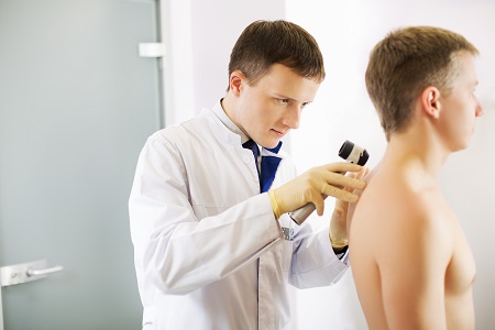
In the early stages, melanoma and skin cancer cannot be seen with the naked eye. Signs of malignant degeneration can only be detected by a specialist using a special device - a dermatoscope. Therefore, dermatoscopy is by far the most effective method of early diagnosis of melanoma and skin cancers.
A dermatoscope is an optical device that takes macro images of skin formations with a 20x magnification. During dermatoscopy, the rays of polarized light penetrate into the upper layers of the skin, which is where the primary accumulation of malignant cells occurs. This information is enough for a dermatologist to assess the condition of the neoplasm and make further recommendations.
In addition to traditional dermatoscopy, the EMC uses the latest digital FotoFinder technology, which combines high-precision dermatoscopy with photographing the entire body and the ability to magnify even the smallest education 140 times. In this way, the entire surface of the skin is examined, all formations on it are analyzed, recommendations for removal are given, and the results are recorded in an electronic map of moles to compare with future examinations.
How is dermatoscopy performed
The procedure is completely painless and comfortable, and lasts no more than 30 minutes.During dermatoscopy, the doctor measures the neoplasm, evaluates the pattern of the edges of the mole, analyzes the surface structure and the degree of penetration of pigmented cells deep into the skin. All moles are recorded in an individual map of skin growths, this is done so that it is possible to track changes over time and make a forecast for the future for observation and removal.
FotoFinder's mole diagnosis also takes 90 minutes and requires no prior preparation. The procedure is fully automated, all moles are automatically entered into the patient's mole chart and analyzed using artificial intelligence. As a result, the patient receives a detailed report with recommendations for removing suspicious moles and caring for the rest. This method improves diagnostic accuracy by up to 99% and allows you to detect even the smallest formations that are invisible during traditional dermatoscopy.
Indications for dermatoscopy
Dermatoscopy is especially recommended:
- patients with light or red hair, fair skin, and light eyes;
- people with a large number of moles, freckles, moles, and age spots;
- elderly people;
- people with a genetic predisposition (in the family there are cases of melanoma and skin cancer);
- patients who spend a long time in the sun;
- patients who have moles that are often subjected to mechanical stress (injured or rubbed by clothing).
Dermatoscopy must be performed before removing the neoplasm, as well as if a new mole has appeared that is growing rapidly, or the old mole has begun to change (asymmetry, peeling, inflammation, itching, bleeding, etc.). In case of injury to the mole or the skin area where it is located.
Dermatoscopy is also used to diagnose:
- keratomas or solar keratosis;
- hemangiomas or angiomas;
- papillomas, warts.
Contraindications
Dermatoscopy is completely safe and has no contraindications. It is recommended for children, pregnant women and women during lactation.
Advantages of dermatoscopy
- The method allows to examine neoplasms of any size
- Does not require surgery, anesthesia, completely painless
- 99% diagnostic accuracy
- It takes no longer than 30 minutes and does not require any preliminary preparation
- The doctor receives the result immediately, and the patient receives recommendations for further examination and treatment
- The method has no contraindications, no complications and no recovery period
- FotoFinder digital dermatoscopy is equipped with a program that automatically issues a conclusion based on the results of the examination, so doctors and the patient can be sure of the accuracy of the recommendations
- Dermatoscopy allows you to track the dynamics of the development of skin tumors
The danger of moles
The World Health Organization uses about 300 names for the term that patients commonly refer to as a mole. Any skin formation can degenerate into melanoma or skin cancer,however, this chance is higher in some formations. In addition, neoplasms can develop on clean skin.
- Dysplastic (atypical) nevi become the most common melanoma. This is a pigmented neoplasm, similar to a mole, with bizarre edges, often of a heterogeneous black-brown color. An atypical nevus may be flat or rise above the rest of the skin in the middle. In the presence of such formations, it is necessary to visit a dermatologist at least once a year. If there are more than 50 of them, screening should be carried out every six months or more often, the doctor will give individual recommendations.
- The probability of becoming melanoma is increased in congenital pigmented nevi (birthmarks). Giant pigmented nevi (more than 10 cm) have the highest risk of degeneration, it is 30%, and they are recommended to be removed as soon as possible.
- Papillomas have the lowest risk of malignancy.
There is a generally accepted system of self-diagnosis
A person can conduct an examination of moles himself, but we must not forget that the accuracy of such screening cannot be compared in accuracy with an examination by a professional dermatologist.
The "ABCDE" rule
"A" (Asymmetry) —Asymmetry. If the mole is no longer round or rounded, it must be shown to a dermatologist.
«B» (Border) — Border.If the edges of the mole have become uneven, jagged or scalloped, this is a reason to consult a specialist.
"C" (Color) is a strong color. Most normal moles are brown or flesh—colored. The appearance of black, red, blue or white shades may indicate the degeneration of a mole into a melanoma.
"D" (Diameter) —Diameter.Most often, moles that have degenerated into melanoma are larger than 6-7 mm.
"E" (Evolving) —Evolution.Any changes that occur with the neoplasm. The most dangerous signs are bleeding, deformity, decrease or increase, the appearance of inflammation, itching, peeling and burning, hair loss from the surface of the mole, the formation of crusts on the surface of the mole, the disappearance of the skin pattern, etc.
If you find at least one of these points, you should consult a dermatologist.
The Ugly Duckling Rule
If a mole has degenerated into a melanoma, it will stand out from the rest of the formations on the skin.This mole needs to be urgently shown to a dermatologist or oncologist.
To detect changes in a timely manner, it is important to regularly conduct a full-body examination. Not forgetting that melanoma can occur not only on the exposed and visible parts of the body, but also on the scalp, behind the ear, on the heels and even on the nails.
Prevention of melanoma and skin cancer
Anyone can get melanoma and skin cancer, but there are people who have an increased risk, they need to follow preventive measures more carefully and strictly follow the individual schedule of examinations and doctor's recommendations. These are people with fair skin, hair, and eyes, with a history of sunburn, a large number of moles, and freckles on their bodies.
General recommendations for prevention:
- Use sunscreens. To protect against the most dangerous ultraviolet A (UVA) and ultraviolet B (UVB) rays on the skin, choose products with an SPF of at least 30 (the higher the SPF, the better) that do not wash off with water. Apply the product to exposed areas of the body, even on a cloudy day. To absorb the protective agent, apply it 20-30 minutes before going out in the sun, repeat the application every 2 hours, especially if you sweat profusely or swim in an open pond. If your cosmetic product contains sun protection components, no additional protection is required. Use sunscreens all year round and do not stay in the sun for more than 2.5 hours.
- Wear long-sleeve shirts and trousers. Wear a baseball cap or a wide-brimmed hat to protect the nose, ears, forehead, and neck area.
- Use sunglasses. Protect your eyes, wear panoramic sunglasses, they block 100% the effects of UVA and UVB rays (glasses must be ANSI approved). The rest of the glasses block only 70% of UV rays, and glasses without special markings may have no protection at all, or have an insufficient level. The degree of darkening of the glass does not matter, protection is provided by treating the glass with special chemicals.
- Plan your day in such a way as to avoid peaks of solar activity and direct exposure to sunlight during periods of maximum activity, from 12:00 to 16:00. Make sure that children play in the shade and are well protected with sunscreen and clothing. Keep in mind that sand, water, and snow reflect UV rays, thereby enhancing their effects. In addition, apply protective measures on cloudy days, as UV rays penetrate clouds perfectly.
- Avoid going to the solarium. Tanning beds and solar lamps emit UVA rays that penetrate deeply into the skin and can provoke the development of skin cancer.
- Perform a regular self-diagnosis. Check for new moles, freckles, and other formations. If you notice changes in existing moles and freckles or see new and suspicious formations, contact a dermatologist.
- Protect yourself from the sun all year round and protect your loved ones. Remember that sunburn can affect the development of skin cancer. Pay special attention to children and adolescents, because it is children's age that can determine the likelihood of developing melanoma and skin cancer in the future.
Advantages of diagnosis and treatment in EMC
- Double control of the results. The possibility of classical dermatoscopy and the innovative FotoFinder method.
- The results of the histological examination are provided as soon as possible thanks to our own histological laboratory.
- All types of mole removal: surgical, laser, electrocoagulation, thermoregulation, chemical.
- Full range of diagnostics: CT, MRI, PET/CT.
- Modern methods of treating melanoma and skin cancer: surgical treatment (sentinel lymph node biopsy), radiation therapy, immunotherapy, targeted therapy.
- The decision on treatment tactics in each case is made by a council of doctors of various specialties. This allows you to correctly determine the stage of the disease and choose the best treatment.
Dermatoscopy is performed in all EMC clinics, including the Children's clinic. FotoFinder can only be completed at the clinic at 35 Shchepkina Street.
Doctors


.jpg)

.jpg)
.jpg)


- Specialization — pediatric and adult dermatology, diagnosis and removal of skin growths in adults and children. He has the skills of dermatoscopy and photographing skin growths
- Graduated from the First Moscow State Medical University named after I. M. Sechenov of the Ministry of Health of the Russian Federation with a degree in Medical Medicine, and completed her residency at the Central State Medical Academy of the Presidential Administration of the Russian Federation with a degree in Dermatovenerology
- Permanent participant of All-Russian congresses and symposiums
