Get help
Specify your contacts and we will contact you to clarify the details.
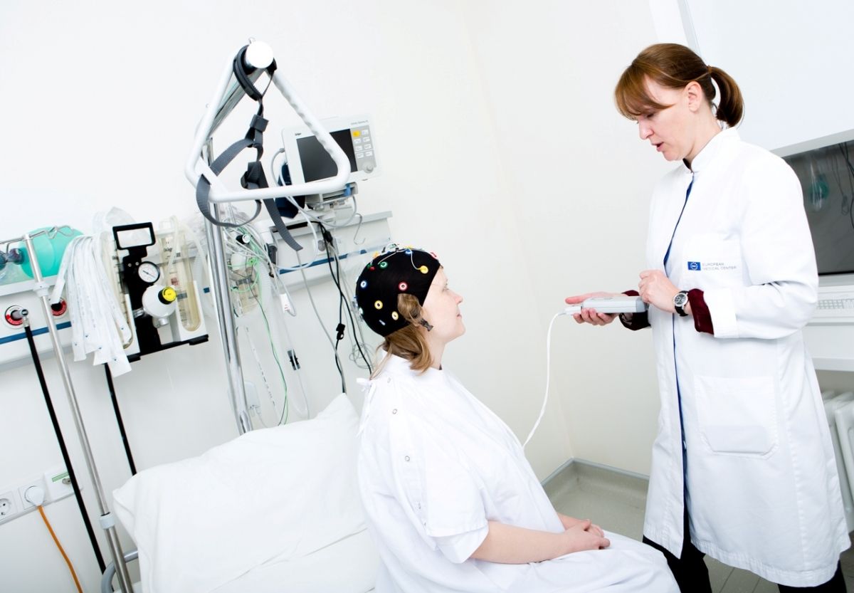
EEG is a method of recording the electrical activity of the brain. The recording process is similar to an electrocardiogram, only the electrodes are not glued to the chest, but to the scalp. During EEG recording, there is no electric shock or medication, only the activity generated by the patient's brain is recorded. The result of the recording looks like a set of curved lines, according to the specific shape of which a neurologist can get information about the work of the brain.
A short EEG lasting several tens of minutes (about 30 minutes on average) is called a "routine EEG." This is a method of rapid diagnosis for suspected epilepsy in the first place. The informative value of a short EEG is not very high, because in a short period it is not always possible to "catch" pathological discharges.
EEG video monitoring is a method of long-term (several hours or days) EEG recording in combination with video recording. In the video, you can see the conditions that bother the patient and determine whether they relate to epilepsy. During the EEG video monitoring, the patient or his relatives accompanying him can indicate the exact time of the onset of seizures using the signal button, this will accurately assess the moment of the appearance of complaints.
EEG monitoring without video recording (long-term EEG monitoring, often lasting a day or more). This study can be performed on an outpatient basis, meaning that after installing the electrode system, the patient leads a normal lifestyle at home or in a hospital.
A doctor or an EEG technician will perform standard tests in which pathological discharges can be seen on the EEG. There are usually three such trials. The first test: the patient is asked to open and close his eyes at a signal. The second test is photostimulation, when a lamp is placed in front of the patient's eyes, flashing at different frequencies. During light flashes, the technician may ask you to look at the lamp, open and close your eyes. The third standard test is hyperventilation, when the patient is asked to breathe deeply and smoothly, usually for 3 minutes (sometimes more or less). Deep breathing can make you feel dizzy, which is normal. For children, hyperventilation can be performed in play when a child blows on a toy (this is usually possible from the age of 3-5) .
With prolonged EEG monitoring, other tests are possible: you can hold your arms outstretched in front of you, repeat the numbers or words mentioned, count from one to one hundred, etc. If a video is being recorded, the technician will show you where the video camera is located and ask you to stay in the camera's field of view during the entire study.
Long-term EEG monitoring is usually performed during wakefulness and during sleep. Most patients do not need to take sleeping pills, as they change the EEG picture. With a study lasting several hours, there is usually enough time to fall asleep on your own. During EEG video monitoring, it is very important to see the patient's movements, including during sleep, so it is important not to cover yourself with a blanket.
When recording an EEG, children always experience movement interference, which is normal. It is more difficult to read an EEG recording with such interference, but in most cases it is quite realistic. It is very rare to have to stop the study because of the child's anxiety, in such cases, you can repeat the EEG on another day. When examining at home without quality control of the recording by the staff, interference is also possible, requiring repetition of the EEG.
After the end of the EEG, the remnants of the gel or paste are wiped off with a napkin and, if necessary, washed off with water. Sometimes the remnants of the paste can be visible on the hair for several hours or days.
Different types of EEG are needed for different purposes. For some patients, a short EEG is sufficient, but sometimes it takes several days to record. The type of examination is determined only by the attending physician.
Usually a neurologist (optimally, a neurologist-epileptologist), less often a psychiatrist. When planning an EEG, it is important to create a research scenario: choose the duration of the study, the number and types of activating samples, the need to record during sleep or during wakefulness, the possibility of reducing sleep duration or discontinuing medications taken by the patient. If you do not know what type of EEG is needed, you should check it with your doctor or discuss it at an appointment with an EMC neurologist-epileptologist.
There are no absolute contraindications.
Relative contraindications : damage to the scalp (wounds, burns, infections), alcohol or drug intoxication, pronounced psychomotor agitation.A hyperventilation test is contraindicated in:
With the listed contraindications to hyperventilation, studies can be performed without a hyperventilation test.
If a prolonged epileptic seizure occurs spontaneously during the EEG or in response to the samples, medical help may be required and the study may be discontinued. If severe seizures are expected to be provoked, it is better to conduct the study in a hospital rather than at home.
The electrodes used to record the EEG can leave visible marks (indentations) on the scalp for up to several hours.
After the end of the study, the patient receives a doctor's report with a description of the EEG and a printout of the indicative sections of the signal ("curves"). If significant events were observed during the video recording study (for example, epileptic seizures), an electronic media (disc) with video clips of seizures is additionally provided. EEG monitoring usually takes time to decode, and the result is given a few days after the examination.
Important! Pathological waves are rarely found on the EEG of healthy people, and vice versa, the patient's EEG may be normal.
It is recommended to discuss the details of the upcoming study with your doctor. If necessary, the doctor can schedule sleep restriction and/or medication withdrawal before the study, and prescribe specific tests.
Children should be told in advance about the need to wear a helmet with wires – playing pilots, astronauts, robots, hairdressers, etc.
Before the study, it is recommended to wash your hair, do not apply styling products to your hair, untangle braids (if available), and remove existing hair clips and jewelry. During a long-term study at the clinic, you can take pajamas with you, which will be comfortable to sleep in without a blanket.
If the patient is agitated and resists putting on the electrode cap, it is acceptable to wait for falling asleep and start the study during sleep. In rare cases, due to the patient's marked resistance, the study may be canceled.
For short studies, it is enough just to sit quietly, follow the instructions of a doctor or an EEG technician.
With EEG monitoring: after the instructions of the doctor / EEG technician and the completion of mandatory activating tests, you can lead a normal lifestyle - eat, drink, read, watch TV, sleep, go to the toilet.