Get help
Specify your contacts and we will contact you to clarify the details.
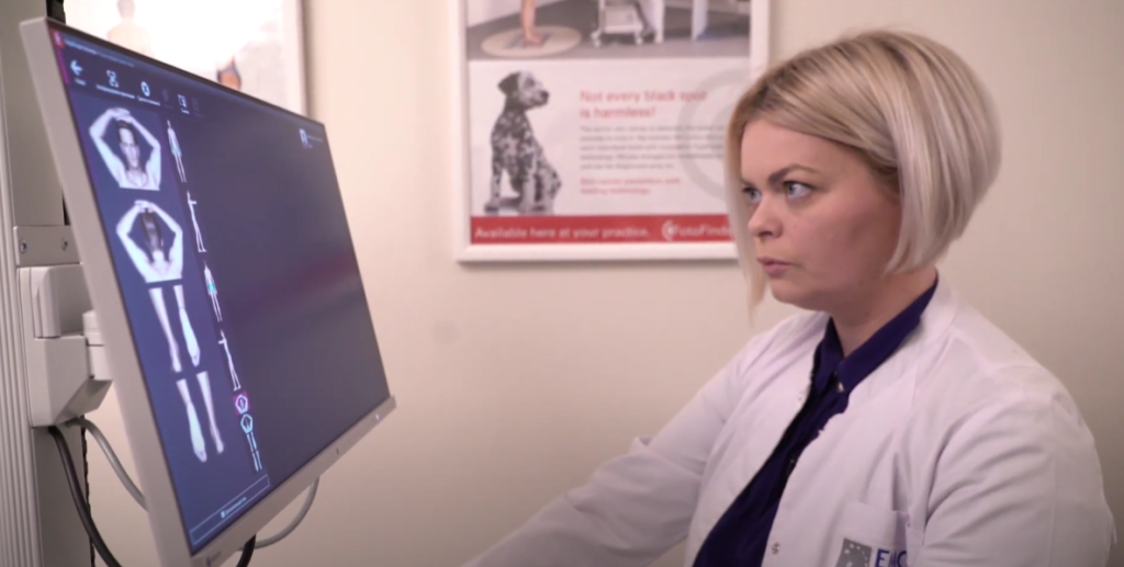
Timely detection of melanoma can be challenging even for an experienced doctor. Recent studies prove that more than 70% of melanomas do not develop from a pre-existing mole, but on "clean skin". At the same time, melanoma at the beginning of its development can be very similar to an ordinary mole, and only a specialist armed with an optical device can suspect this dangerous skin formation. Early detection of melanoma is the main factor in its successful treatment. Therefore, the most effective solution for early diagnosis of dangerous pathology is to create a "mole map" and regular dermatological screening of the entire skin surface, which will identify any neoplasm. The EMC clinic uses the innovative FotoFinder ATBM (Automatic Total Body Mapping) diagnostic system for this purpose.
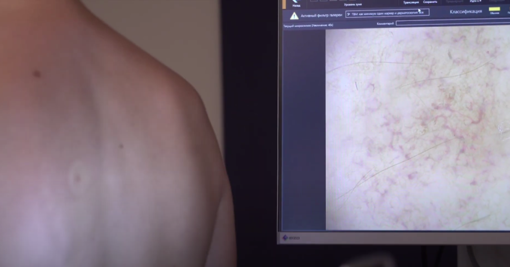
FotoFinder ATBM is a hardware and software package that photographs the entire surface of the skin, then a dermatologist performs a dermatoscopy and analyzes the detected neoplasms, performing digital mapping of the detected moles. Dermatoscopic images of skin formations are automatically stored in computer memory, which allows them to be further analyzed and compared during the observation process.
The hardware part of the diagnostic complex is represented by a powerful modern video dermatoscope Medicam 1000. This is a device equipped with a Full HD video camera and a high-quality lens with optical magnification up to 400x.
The software part of the FotoFinder ABTM system is represented by the following technologies:
The FotoFinder software archive is, in fact, a worldwide medical library that stores dermatological data from patients around the world. The program's interface provides a second opinion service — a dermatologist can request advice from world-class experts who also use the FotoFinder diagnostic complex at any time.
In addition to detecting malignant neoplasms, the examination provides important diagnostic information in the treatment of other dermatological pathologies: microvascular abnormalities, parasitic invasions, dermatoses, psoriasis, etc.
Skin cancer is one of the most common types of oncopathologies. Melanomas account for up to 3% of all detected cancer cases, and the risk of getting sick for a person of Caucasian race exceeds 2.5%. At the same time, melanoma accounts for up to 80% of deaths in the group of skin malignancies, however, with the detection of a tumor at an early stage and timely treatment, the survival rate almost reaches 100%. Therefore, we recommend digital dermatoscopy on the FotoFinder complex to all patients who care about their health.
The study is safe, painless and can be performed in both adults and children. Preferably, monitoring with this device in adults and adolescents who have completed growth, since the computer will be able to more accurately compare the results with the data from the previous survey. Nevertheless, research using the Fotofinder system may also be useful for children. Digital mapping does not require special preparation or anesthesia. The duration of the procedure ranges from 30 to 90 minutes, depending on the number of nevi on the patient's skin. For the procedure, the patient will need to take off his clothes so that the digital mapping is as complete as possible.
Stages of the procedure:
The FotoFinder diagnostic complex allows you to detect skin diseases at a pre-symptomatic stage with an accuracy of up to 99%. A digital map of moles is the best way to diagnose dangerous pathologies early. Make an appointment for an examination online or by phone +7 495 933-66-55.