

Biopsies under the control of ultrasound, CT, MRI
Biopsies under the control of ultrasound, CT, MRI
Puncture biopsy under the control of CT, MRI or ultrasound is a minimally invasive percutaneous procedure that allows to obtain histological material and avoid diagnostic surgery.
At the EMC Institute of Oncology ultrasound-controlled biopsies are performed by an experienced specialist in ultrasound diagnostics with the presentation of material for a complete pathomorphological examination.
Ultrasound-controlled biopsies are performed when the tumor is localized in the peripheral lymph nodes, thyroid gland, liver, kidneys and other organs.
In cases where a biopsy under ultrasound control is not possible, needle biopsies are performed under CT control. CT-controlled biopsies are performed with deeply located formations, in particular, formations in the lymph nodes of the mediastinum and retroperitoneal space, pancreas, lungs, as well as tumors of the kidneys, adrenal glands, liver, and pelvis.
Biopsies from formations in the lungs allow obtaining material for a complete pathomorphological examination, as well as the genetic characteristics of the tumor for targeted treatment.
CT-controlled biopsies are performed by an international radiological diagnostic specialist Professor Evgeny Libson, who has experience in conducting more than 12,000 puncture biopsies.
MRI-controlled biopsies of breast formations are performed in cases where these formations are not visible on mammography and ultrasound, but visible only on MRI.
The experience of the first breast MR biopsy in Russia belongs to the staff of the European Medical Center. Currently, this type of biopsy is also standard and routine and does not require prior preparation.
Why the EMС
The first and only clinic in Russia, created in the image of the world's leading clinics
EMC is a multidisciplinary center offering patients a high level of medical services and a personalized approach
600
world-renowned doctors
57
treatment directions
36
years taking care of your health
24/7
we work at any convenient time
World recognition and awards
Our achievements are confirmed by prestigious international awards
 More details
World recognition and awards
We work according to international standards, we have licenses and certificates
 Certificates and licenses
Get help
Specify your contacts and we will contact you to clarify the details.
Doctors

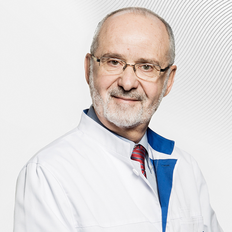
Evgeniy Libson (Israel)
Deputy Director of the Institute of Oncology, Chief Consultant on Cancer Diagnostics, Professor of Radiology. Chief Specialist in Cancer Diagnostics and CT-guided Biopsies, FRCR, FRCR
-

Kirillova Margarita
Ph.D. of Medical Sciences, M.D.
-

Trebushenkov Andrey
Head of the Department of Radiation Diagnostics of the EMC, specialist in biopsies under the control of CT
-
Evgeniy Libson (Israel)
Deputy Director of the Institute of Oncology, Chief Consultant on Cancer Diagnostics, Professor of Radiology. Chief Specialist in Cancer Diagnostics and CT-guided Biopsies, FRCR, FRCR
- Actively participates in the work of scientific and research institutions
- He currently holds the position of Professor of Radiology at Hadassah Hospital. Member of the Education Committee of Hebrew University – Hadassah School of Medicine
- Awarded for outstanding contribution to Israeli Healthcare - Israeli Medical Association
Total experience
54 years
Experience in EMC
since 2011
Reviews
Anonymous,
City: -
Super nice
Same as 1st visit Welcome and Secretary were super nice Doctor Abdulin
was great and super pro.
Clinic:
Institute of Oncology
Doctor:
Iskander Abdullin
3 July 2025
Anonymous user Name,
City: -
I wish you all good health and professional success
Many thanks to the doctors Moskalets E. R. and Penkova O. V., who described
my pictures on the day of the study, as well as to the staff of the PET CT department Nikolay, nurse Ekaterina, laboratory assistants Eduard and Nikolay, who were with me from the beginning and until the
end of the study, we worked in a coordinated, professional and friendly manner.
... more
Clinic:
Institute of Oncology
Doctor:
Elina Moskalets
26 March 2025
Konstantin Vladimirovich,
City: -
I express my gratitude for the treatment!
He came back with severe pain in the neck. The diagnosis revealed an intervertebral
hernia. The very next day, the operation was carried out, everything was very fast. After the operation, he was surrounded by nurses. I would especially like to express my gratitude to:
Krivoshapkin Alexey Leonidovich, Yulia Markina, Vladimir Klimov, Orkhan Abdullayev, Gleb Sergeevich Sergeev, and Alexander Loginov.
... more
Clinic:
Institute of Oncology , Neurosurgery Clinic
Doctor:
Krivoshapkin Alexey
16 March 2025
Anonymous user Name,
City: -
We thank all the staff of the clinic for saving our lives
We thank Alexey Leonidovich Krivoshapkin, Alexey Sergeevich Gaitan, Orkhan
Abdullayev and all the clinic staff for saving our lives.
Clinic:
Institute of Oncology , Neurosurgery Clinic
Doctor:
Krivoshapkin Alexey
7 March 2025
Anonymous user Name,
City: -
Endless gratitude
Endless thanks to my dear Dr. Pavel Koposov.
Clinic:
Institute of Oncology
Doctor:
Pavel Koposov
4 March 2025
Vladimir,
City: Yessentuki
About surgeon Ruchkin D. V.
Dmitry Ruchkin is the epitome of the highest professionalism , dedication,
modesty and dedication to his work. His method of gastroectomy is unique, allowing you to eat and live a full life after a complex operation. A low bow to him and the surgeon Jan to Maria Nikolaevna —
members of his team.
... more
Clinic:
Institute of Oncology
Doctor:
Dmitry Ruchkin
12 February 2025
Anonymous user Name,
City: -
Thank you to all the clinic staff!!!
All employees throughout the chain from reception to the end of the procedure
were very kind and attentive. I would like to express my gratitude to all of you, and I would like to especially mention doctor Ilya E. Loiko for his kind words and wishes. Thank you to all the clinic
staff!!!
... more
Clinic:
Institute of Oncology
Doctor:
Ilya Loyko
24 January 2025
S. V. Firmanov,
City: -
Gratitude from the patient
Many thanks to all the staff of the clinic, especially Pavel Koposov.
Clinic:
Institute of Oncology
Doctor:
Pavel Koposov
16 January 2025
Anonymous user Name,
City: -
Many thanks to the professional in his field
Thank you to the team of the Center for Radionuclide Diagnostics for their
well-coordinated, productive work and good mood during the preparation and conduct of the study. And, of course, a huge thank you to the professional in his field, Dr. Borzyanitsa. Happy New Year Happy New
year to the entire EMC team!
... more
Clinic:
Institute of Oncology
Doctor:
Stanislav Borzyanitsa
25 December 2024
Anonymous user Name,
City: -
Gratitude
Sincere thanks to Dr. Vasilyeva.
Clinic:
Institute of Oncology
Doctor:
Irina Vasilieva
12 December 2024
Anonymous user Name,
City: -
Thank you all very much from me and my family
Many thanks and best wishes from the bottom of my heart: Zhao V. A., Yuldashev
A. G., Vakhabova Yu. V., Zhizhko N. V. specialists from GOD. The medical staff is beyond praise.
Clinic:
Institute of Oncology
Doctor:
Anvar Iuldashev
12 December 2024
Anonymous user Name,
City: -
Thank you very much
Many thanks to Iskander Abdullin, the staff of the Center for Radiation
Diagnostics.
Clinic:
Institute of Oncology
Doctor:
Iskander Abdullin
11 December 2024
Anonymous user Name,
City: -
You can trust such professionals
The most important thing is care and tact at all stages of visiting the
clinic and, as a conclusion, professionalism of the highest level Dr. Irina Vasilyeva, who surprisingly combines knowledge, skills, experience, and sensitivity and empathy.
Clinic:
Institute of Oncology , Breast Clinic
Doctor:
Irina Vasilieva
9 December 2024
Anonymous user Name,
City: -
Gratitude to the doctor
Thanks to the doctor Vakhabova Julia Vyacheslavovna, she blessed me for
6 months...
Clinic:
Institute of Oncology
Doctor:
Yuliya Vakhabova
28 November 2024
Anonymous user Name,
City: -
I express my deep gratitude to the specialists of the highest class
I would like to express my deep gratitude to the top-class specialists,
Professor Yevgeny Libson, Director of the Cancer Center Salim Nidal, and Ilya Yevgenyevich Loiko for my essentially "salvation", for the extremely kind and sincere attitude.
Clinic:
Institute of Oncology
Doctor:
Evgeniy Libson (Israel)
26 November 2024
Anonymous user Name,
City: -
I got ALL my questions answered
I was at the consultation of Professor Vsevolod Borisovich Matveev... I
got ALL my questions answered... And even more...In a word-not a man, but a HUMAN BEING...!!! The staff is friendly, answers all questions, helps, etc... People with health problems come in...They all have no
time for this or that...Employees understand all this and make their stay in the clinic as comfortable as possible...
... more
Clinic:
Institute of Oncology , Urology Clinic
Doctor:
Vsevolod Matveev
22 November 2024
Popov Khariton,
City: -
Thank you very much
Many thanks to Iskander Abdullin, Alexey Krivoshapkin, Natalia Zharinova,
Elmira Sultanova, Alla Artamonova and all the staff of the clinic who helped me and my children.
Clinic:
Institute of Oncology
Doctor:
Iskander Abdullin
20 November 2024
Anonymous user Name,
City: -
Words of gratitude to A. L. Krivoshapkin
Special thanks to Professor A. L. Krivoshapkin for the online consultation.
And I want to thank the girl operator who helped me with installing the app for the conference.
Clinic:
Institute of Oncology , Neurosurgery Clinic
Doctor:
Krivoshapkin Alexey
31 October 2024
Anonymous user Name,
City: -
Thanks!
The EMS clinic was contacted on the recommendation of a friend of my husband.
Thank you for your cordiality, care, and attention from the first visit to the clinic, starting with the staff meeting in the lobby, reception, and further support throughout the entire examination,
treatment options.I would like to thank the Oncology department , my attending physician Stanislav Borzyanitsa (for his attention, care, cordiality, support during treatment...)I would like to thank DIO Salim Nidal, the staff, the staff of the Radiotherapy Center, Radionuclide Diagnostics, performing a biopsy, the Staff of the Hospital and Laboratory...Thanks!
... more
Clinic:
Institute of Oncology
Doctor:
Salim Nidal (Israel)
30 October 2024
Anonymous user Name,
City: -
Sincerely grateful
I am sincerely grateful to Dr. Salim Nidal, Dr. Libson, Dr. Anatoly Olegovich,
Valyusha and others, as well as to the girls at the reception and the entire radiotherapy team. Thank you very much for your professional, attentive and warm attitude. Strength to you and health!May God
grant that we will work for many more years and bring JOY to people!
... more
Clinic:
Institute of Oncology
Doctor:
Salim Nidal (Israel)
29 October 2024
Licenses and certificates
The EMC's activities comply with internationally accepted medical and administrative standards, as well as patient safety requirements.
ICI INTERNATIONAL QUALITY CERTIFICATES ISSUING SERVICES LLC
International Accreditation
Joint Commission International
International Accreditation 2018, 2021
Luxury Lifestyle Awards
Best Luxury International Private Hospital Group
International Hospital Federation
International Hospital Federation Premier Member
IV All-Russian rating of radiology departments
Four-time winner
License for medical activities
License for pharmaceutical activities
License for activity on circulation of narcotic drugs, psychotropic substances
License for providing high-tech medical care