Hysteroscopy of the uterus
- Diagnostic and surgical hysteroscopy for endometrial polyps, fibroids, adhesions, and other problems.
- Hysteroscopy using an ultra-thin device, safe for use in nulliparous patients.
Uterine diseases are detected, according to WHO, in 40% of women. It is often these pathologies that cause female infertility.
To identify the pathology and safely remove neoplasms of the uterus, the specialists of the European Medical Center use an effective modern technique – hysteroscopy.
The procedure allows you to assess the condition of the uterine cavity and detect pathology even at an asymptomatic stage of development.
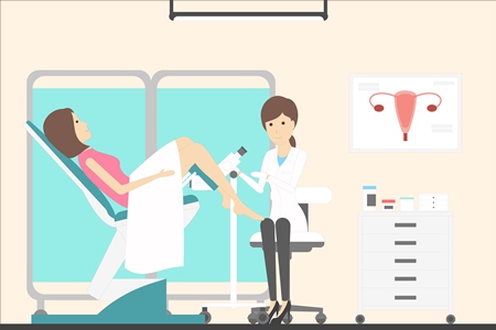
How is hysteroscopy performed
After treatment with an antiseptic solution of the external genitalia, a hysteroscope is inserted into the uterine cavity, a special optical instrument that transmits an image magnified several times (up to 10) onto the screen. The doctor examines the cervical canal, the uterine cavity, and the mouth of the fallopian tubes. During the examination, the gynecologist evaluates the thickness, uniformity and color of the endometrium, and also checks for polyps and submucosal myomatous nodes.
Hysteroscopy can be diagnostic or surgical. Diagnostic is performed to make a diagnosis and to monitor after surgical treatment. It is performed without anesthesia or with the use of local anesthesia.
During surgical hysteroscopy, it is possible not only to identify adhesions or neoplasms in the uterus, but also to remove them. The removed tissues are then sent to a laboratory for histological analysis. Short-term intravenous anesthesia is used for surgical hysteroscopy.
Indications for hysteroscopy
Indications for diagnostic hysteroscopy:
- suspected endometrial disease (hyperplasia/polyps) detected during ultrasound;
- suspicion of a submucosal node;
- infertility. Suspected internal endometriosis, synechia in the uterine cavity;
- malformations of the uterus; removal of a foreign body (for example, an intrauterine device).
Indications for operative hysteroscopy:
- submucosal fibroids (hysteroscopic myomectomy);
- intrauterine septum;
- hyperplasia, polyps, endometrial cancer (polyp resection, RV);
- removal of a foreign body;
- adhesions and synechia (hysteroscopic resection);
- abnormal uterine bleeding.
The need for a study is determined only by a doctor.
Safety of hysteroscopy
Hysteroscopy is a safe and easily tolerated procedure. You can leave the clinic within an hour or two after the manipulation.
In the presence of endometrial neoplasms that interfere with pregnancy, hysteroscopy successfully solves the problem.
However, complications cannot be excluded, which in exceptional cases may occur during the procedure:
- Acute inflammatory diseases of the pelvic organs;
- If breast cancer is performed during hysteroscopy, a possible complication may be the development of scar tissue in the uterine cavity - Ascherman's syndrome.
How to prepare for hysteroscopy
The procedure is usually performed on the 6th-10th day of the menstrual cycle.
It is recommended to refrain from sexual intercourse 2 days before the study, and from douching and using tampons a week before.
If you are taking any medications in the form of vaginal suppositories, pills, etc., you should tell your doctor about this.
Preparation for office hysteroscopy:
-
laboratory tests: clinical analysis of blood, urine, hospital complex, ECG, smear on flora.
Preparation for operative hysteroscopy:
-
laboratory tests: clinical analysis of blood, urine,hospital complex, biochemical blood test, coagulogram. ECG, smear on flora;
- consultation with an anesthesiologist, therapist;
- chest X-ray examination.
What to expect after the procedure
After an office hysteroscopy, the patient can immediately be free, minimal genital discharge is possible - this is the norm.
After an operative hysteroscopy, the patient spends several hours in the ward to recover from anesthesia and observe the general condition and volume of spotting. A few days after the procedure, in addition to discharge, there may be minor pulling pains in the lower abdomen. During 1-2 weeks after surgery, it is necessary to refrain from sexual intercourse, taking baths, swimming pools, saunas, and using vaginal tampons.
Contraindications to hysteroscopy
- A desirable pregnancy;
- Infections of the pelvic organs.
Advantages of contacting the EMC
-
Safety: manipulation is performed with an ultrathin hysteroscope with a diameter of 3-5 mm, which does not require dilation of the cervical canal. This is especially important for unborn patients.
- High efficiency: during hysteroscopy in the EMC, it is possible to undergo atraumatic removal of uterine cavity polyps or submucous myomatous nodes using a hysteroresectoscope with simultaneous coagulation of the pedicle. This eliminates the recurrence of the disease in almost 100% of cases.
- Doctors with work experience in leading medical centers in Europe and the USA. Diagnosis and treatment according to international protocols.
- Interdisciplinary approach: we organize consultations with the participation of the necessary specialists (gynecologist, reproductologist, endocrinologist, oncologist, etc.).
Why the EMС
The first and only clinic in Russia, created in the image of the world's leading clinics


Get help
Specify your contacts and we will contact you to clarify the details.
Doctors

Evgeniy Libson (Israel)
Deputy Director of the Institute of Oncology, Chief Consultant on Cancer Diagnostics, Professor of Radiology. Chief Specialist in Cancer Diagnostics and CT-guided Biopsies, FRCR, FRCR
-

Salim Nidal (Israel)
Director of the Institute of Oncology
-

Vladimir Nosov
Ph.D. of Medical Sciences
-

Ron Schonman
Doctor of Medicine
-

Boris Sakhnovich
Doctor of Medicine
-

Dmitriy Subbotin
Ph.D. of Medical Sciences
-

Keburija Lela
Ph.D. of Medical Sciences
-

Nenahov Filipp
Doctor of the highest category
-

Mskhalaya Maria
Ph.D. of Medical Sciences
-

Desyatkova Nina
-

Belousova Nadezhda
-

Saakyan Gayane
Doctor of the first category
-

Shpachenko Viktoria
-

Borovkova Ekaterina
Doctor of the highest category, Professor, Doctor of Medicine
-

Madan Korneliya
-

Kovaleva Larisa
Specialist in Gynecological Endocrinology, Ph.D. of Medical Sciences
-

Loginova Olga
Ph.D. of Medical Sciences
-

Charkhifalakyan Arevik
Head of the Gynecology and Oncogynecology Clinic, Ph.D. of Medical Sciences
-

Panfilova Olga
Leading specialist in prenatal fetal diagnosis, Ph.D. of Medical Sciences
-

Rivkina Natalya
Consultant psychotherapist from Israel, Israeli license number 1175123, Ph.D. of Medical Sciences
-
Evgeniy Libson (Israel)
Deputy Director of the Institute of Oncology, Chief Consultant on Cancer Diagnostics, Professor of Radiology. Chief Specialist in Cancer Diagnostics and CT-guided Biopsies, FRCR, FRCR
- Actively participates in the work of scientific and research institutions
- He currently holds the position of Professor of Radiology at Hadassah Hospital. Member of the Education Committee of Hebrew University – Hadassah School of Medicine
- Awarded for outstanding contribution to Israeli Healthcare - Israeli Medical Association
Total experience
54 years
Experience in EMC
since 2011