MRI of the heart and blood vessels
The EMC Heart and Vascular Clinic performs a wide range of heart examinations using magnetic resonance imaging and computed tomography. The research is carried out by highly qualified doctors with dual specialization in the field of radiation diagnostics and cardiology.
Magnetic resonance imaging (MRI) of the heart is a method for assessing viable myocardium and myocardial blood supply. Magnetic resonance angiography (MR angiography) is a highly informative and safe method for obtaining vascular images.
Magnetic resonance imaging is performed using the Siemens Magnetom Aera tomograph, which opens up tremendous opportunities for obtaining high-resolution, wide-field images in any plane. The large diameter of the CT scanner tunnel window (70 cm) and its short length make it possible to conduct research in patients with claustrophobia (fear of enclosed or cramped spaces).
MRI of the heart and blood vessels in EMC
- MRI of the heart
- MR angiography of head and neck vessels
- MR angiography of the abdominal aorta and its branches with intravenous contrast
- MR angiography of the renal arteries with intravenous contrast
- MR angiography of the arteries of the lower extremities with intravenous contrast
- Contrast-free MR angiography of pelvic and lower extremity arteries
The main indications for MRI of the heart:
-
IHD is an ischemic heart disease (it allows you to assess the volume parameters, contractility of the myocardium, as well as the degree of myocardial damage).
- Cardiomyopathy – changes in the structure and function of the heart muscle (hypertrophic, dilated, ischemic, etc.). Here, MRI allows you to assess the structure of the heart muscle even without using contrast agents.
- Myocarditis is an inflammation of the heart muscle. MRI allows you to visualize areas of extracellular water. After intravenous contrast, the drug selectively accumulates in places where extracellular water accumulates, thereby changing the resonant properties of tissues. Thus, it is possible to accurately determine the localization and extent of the inflammatory process in the heart muscle.
- Diseases of the pericardium (the outer lining of the heart). MRI allows you to accurately assess the structure of the pericardium, determine its thickening, the presence of calcification areas in it; effusion (accumulation of fluid) in the pericardial cavity and bulky formations.
- Acquired heart defects. It allows you to evaluate the function of the heart valve apparatus (to identify regurgitation (reverse blood flow), to estimate its area, and also to obtain a three-dimensional model of the heart valve), to estimate the blood flow rate.
- Congenital heart defects. Assessment of intra- and extracardiac anatomy, often complex combined heart defects; determination of septal defects, intracardiac discharge, etc.
- Tumors of the heart: they can be pseudotumor (parietal and valvular thrombi) and truly tumor (myxoma, rhabdomyoma, fibroids, lipomas, teratomas, etc.).
Get help
Specify your contacts and we will contact you to clarify the details.
Doctors
.jpg)
Plakhova Victoria
Doctor of Medicine
-

Anikeva Evgeniya
Head of the Hospital of the Department of Cardiology and X-ray Endovascular Methods of Diagnosis and Treatment
-

Burakova Natalya
Doctor of the highest category, Doctor of the highest category
-

Dyagileva Mariya
Doctor of the highest category
-

Ignateva Oksana
Head of the Heart and Vascular Clinic, Ph.D. of Medical Sciences
-
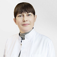
Plakhova Victoria
Doctor of Medicine
- Currently, he is the Head of the Ultrasound Diagnostics Department at the National Research and Practical Center for Cardiovascular Surgery named after Bakuleva" Ministry of Health of the Russian Federation
- Graduated from the Russian State Medical University with a degree in Medical Science
- She completed her academic residency in Cardiology at the A.N. Bakulev Scientific Center for Cardiovascular Surgery of the Russian Academy of Medical Sciences
Total experience
29 years
Experience in EMC
since 2017


