Computed tomography (CT)- X—ray examination based on obtaining layered images of internal organs. It is an accurate, fast and painless method of radiation diagnosis.
The
brain is the only organ in the human body that is almost completely surrounded by bone, so it is impossible to assess its condition without the use of radiation research methods.
Computed tomography of the brain also shows whether there are disorders of cerebral circulation.
In addition, CT scans of the brain are performed in patients who are contraindicated for MRI, namely patients with vascular clips, aneurysm embolization components, certain types of pacemakers, and other devices containing metal
.
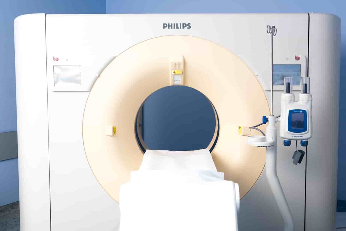
The European Medical Center has the most advanced diagnostic equipment to date. The IDose software package, which is equipped with a Philips i256 multispiral computed tomograph, allows you to obtain high-quality images of the brain with minimal radiation exposure. The clinic uses modern research protocols (according to the latest recommendations of the American and European Radiological Communities (ESR, RSNA)).
High—precision equipment and international research protocols are not the only reasons why brain CT scans should be performed at the European Medical Center in Moscow.
Among other advantages of the EMC:
- the department of radiation diagnostics works around the clock, without days off and holidays, the study can be performed at any time convenient for you;
- most studies are analyzed independently by two specialists (the staff has a specialist in neuroradiology who advises all studies of the central nervous system conducted in the department);
- particularly difficult cases are submitted for discussion by a council of doctors (in this case, by a separate council of doctors specializing in head and neck diseases);
- The head of the EMC Radiation Diagnostics Department is Professor Evgeny Isaakovich Libson (Israel), Honorary Member of King's College (Great Britain), member of the European and North American Radiological communities, the most experienced specialist in radiation diagnostics;
- All research data is uploaded to the PACS electronic medical system, which allows doctors to view images from anywhere in the world and using any electronic device and give their comments and recommendations
The impeccably polite and qualified administrative staff ensures maximum comfort for patients.
CT scan of the brain with contrast
To improve the visibility of brain structures and more accurately determine the presence of a pathological process, computed tomography of the brain with intravenous contrast is used.
Contrast is also used in CT angiography of the brain. This study is conducted to assess the vascular bed, the anatomy of the blood supply to the brain, as well as to identify pathological changes in the cerebral vessels.
The need for intravenous administration of a contrast agent and the main limitations in conducting CT scans of the brain are related.
If you have:
- severe allergic reaction to contrast agent;
- allergic reactions to food, medications, or bronchial asthma;
- diseases of the thyroid gland;
- kidney failure;
- severe form of diabetes mellitus (diabetic angiopathy, nephropathy, etc.),
the EMC will give you recommendations on drug preparation for the study, prescribe tests if necessary, and if it is impossible to perform a CT scan of the brain with contrast, they will select an alternative, a safe research method for you.
Preparation for the study

No training is required for computed tomography of the brain without intravenous contrast.
For conducting this study with intravenous contrast:
- to clarify the presence of contraindications for the administration of a contrast agent;
- elderly patients, patients with known kidney diseases or undergoing chemotherapy, we recommend taking a blood test to determine the concentration of creatinine in the blood (determination of the filtration capacity of the kidneys);
- in the presence of allergic reactions or a moderate decrease in the filtration capacity of the kidneys, carry out drug preparation for the study
- Do not eat less than 2 hours before the study.
- if you are taking tablet forms of sugar-lowering drugs, we ask you to notify the radiologist about this.
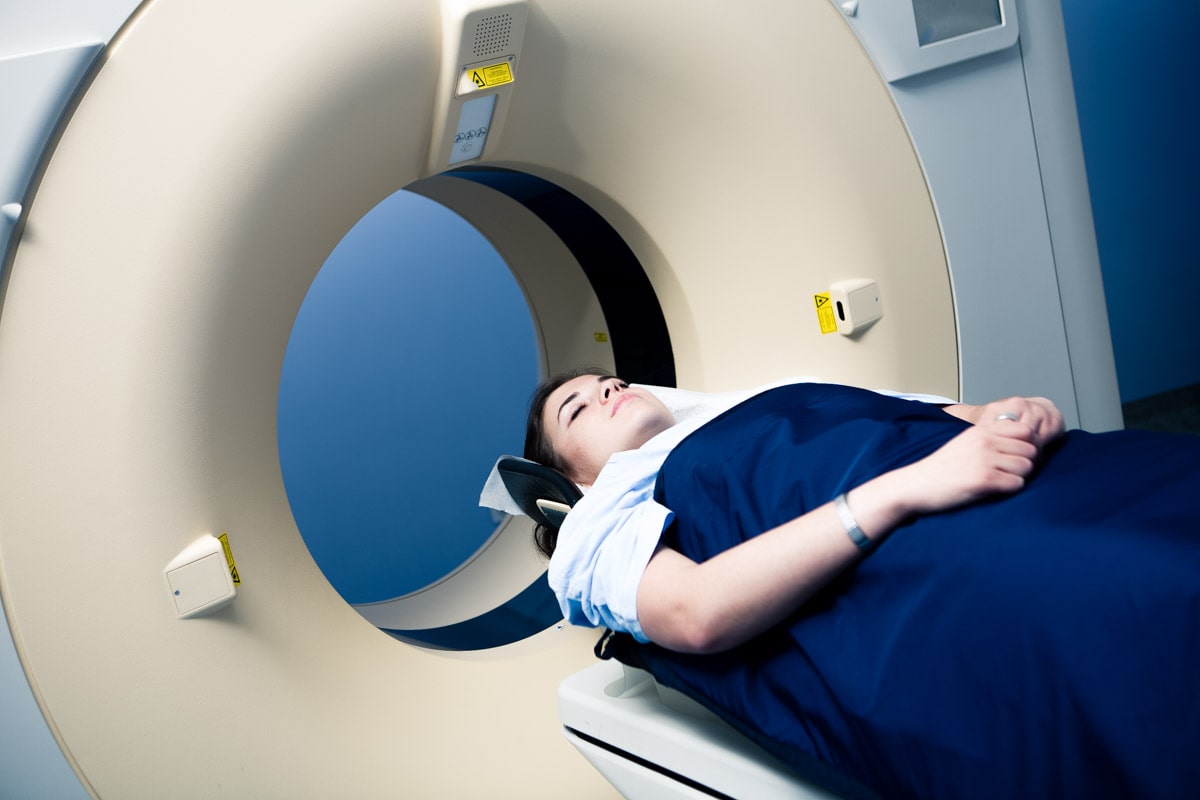
The procedure for conducting the study
If you have previously undergone any research, and you have the results in your hands, you can bring them with you on any carrier - this will help doctors assess the dynamics of the disease..
Before the examination, it is necessary to remove metal objects that fall into the scanning area. Next, the X-ray technician will explain to you the rules of conduct during the examination and will give further instructions while in the control room. The scanning process itself takes a few seconds, and the entire procedure (with changing clothes and contrast agent) will take no more than 15 minutes.
The images obtained as a result of the study are processed by a computer and transferred to professional workstations of radiologists. The final conclusion will be drawn up based on the results of the study by specialists.
The
results of the study are the radiologist's conclusion and the data from the study itself.
You can receive them in any form convenient for you:
—conclusion: original on paper or a copy in electronic form,
—research data: on disc, via link to view in electronic form or, if necessary, onthe film.