Addresses of clinics:
- Moscow, Prospekt Mira metro station, Shchepkina str., 35
- Moscow, Prospekt Mira metro station, Orlovsky lane, 7

Computed tomography of the paranasal sinuses is an accurate, non-invasive and safe diagnostic examination. The method is based on the use of X-ray radiation. Unlike radiography, CT scans produce a series of images that can be transformed into a 3D image.
In the radiology departments of the European Medical Center in Moscow, you can undergo round-the-clock computed tomography of the sinuses using the most modern equipment available today – the Phillips multispiral computed tomographiCT 256, equipped with the iDose software package, which significantly reduces radiation exposure. Your research will be independently analyzed by two highly qualified specialists, after which you will be given a conclusion in any form convenient for you.In the EMC clinics, you will find unsurpassed quality of medical care, comfort and convenience inherent in the best clinics in Western Europe, the USA, and Israel.
The main indications for computed tomography of the paranasal sinuses are:
- suspected presence of various diseases of the nose and paranasal sinuses;
- suspected diseases of the middle and inner ear;
- injuries to the facial skeleton;
- planning of reconstructive plastic surgery in the area of the facial skeleton.
In most cases, computed tomography of the paranasal sinuses is performed without intravenous contrast and does not require special training. Therefore, the only limitation of this method may be pregnancy. However, taking into account the use of modern hardware and software and if there are objective clinical indications, this study does not pose a danger to pregnant patients.
If the clinical situation still requires intravenous contrast (for example, if cancer of the nasal mucosa is suspected), then there will be contraindications associated with the use of a contrast agent containing iodine (allergic reactions, renal and heart failure, thyroid diseases, severe forms of diabetes mellitus).
In each specific case, the need for a contrast agent is determined by the radiologist.
How is the CT scan of the sinuses
Before the examination, you will be asked to remove metal objects that fall into the scanning area. The X-ray technician will tell you how the study will take place and answer your questions.Computed tomography of the paranasal sinuses takes no more than 10 minutes, including changing clothes. The scan itself lasts a few seconds.
Results of a CT scan of the paranasal sinuses
After the study, the images obtained are processed by a computer and transferred to the professional workstations of our radiologists. Most of the studies in the department are analyzed independently by two specialists, and, if necessary, are submitted for discussion within the framework of regular consultations of doctors specializing in diseases of the head and neck organs.
What does a CT scan of the paranasal sinuses look like?
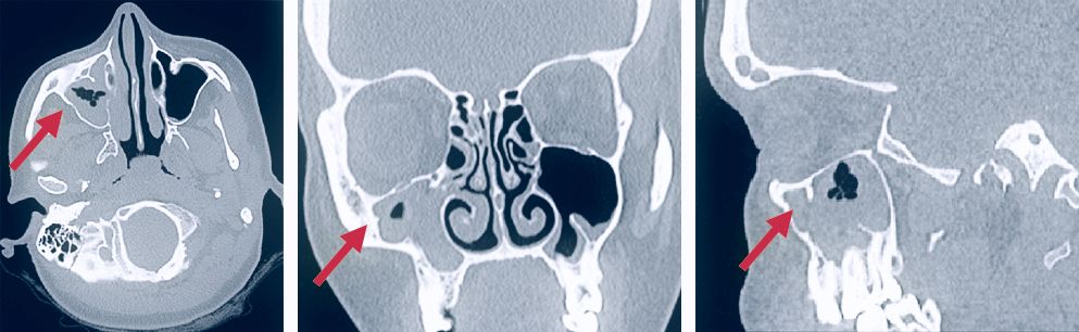
The arrows in the image indicate the pathological contents in the right maxillary sinus.
This is how the radiology department doctor sees your study. Based on the images obtained, he will be able to assess the anatomy of the paranasal sinuses and other organs of the area, diagnose the disease and help the clinician choose the optimal treatment regimen.
Conducting research for children
To obtain clear images during computed tomography of the sinuses, the patient's immobility during the examination is important. Children are a category of patients who find it difficult to stay motionless even for a short period of time. Therefore, in the EMC, such studies can be performed under medical sleep, in the presence of an experienced anesthesiologist. In addition, one of the parents will be able to stay with the child during the study. It is only necessary to wear an apron that protects against X-rays.
What is the difference between CT and MRI?
This is the most common question that is asked to a radiologist. And this is not accidental, because the devices are very similar in structure – they are a structure with a tunnel in the center and a table that drives into the tunnel. The tunnel of an MRI scanner is longer, and being in it is less pleasant for people with a fear of confined spaces. However, this is far from the only difference. The most important thing is that the principle of image acquisition is completely different.
CT uses X-ray radiation, and images are constructed based on varying degrees of absorption of this radiation by tissues.
MRI uses a magnetic field. The image is based on the physiological structure of organs and tissues and depends on the number of water molecules in the body tissues.
In modern medicine, there is no single examination technique that could provide answers to all the questions that arise. All methods of radiation diagnostics complement each other.
Only qualified doctors of clinical specialties, together with radiologists, can correctly prescribe radiation examinations, correctly interpret their results and formulate an accurate diagnosis.