Addresses of clinics:
- Moscow, Prospekt Mira metro station, Shchepkina str., 35
- Moscow, Prospekt Mira metro station, Orlovsky lane, 7
Computed tomography of the kidneys allows you to obtain their diagnostic images, including in 3D format. This study is used for early detection of pathological processes in the kidneys and adrenal glands.
Kidney CT is usually prescribed for:
- suspected congenital anomalies;
- suspected presence of concretions (stones)in the urinary tract;
- assessment of the density of kidney stones before the crushing procedure;
- injuries of the kidney area;
- diagnostics of neoplasms and metastases;
- kidney health monitoring after surgery;
- determining the site for taking a biopsy;
inflammatory and other kidney diseases.
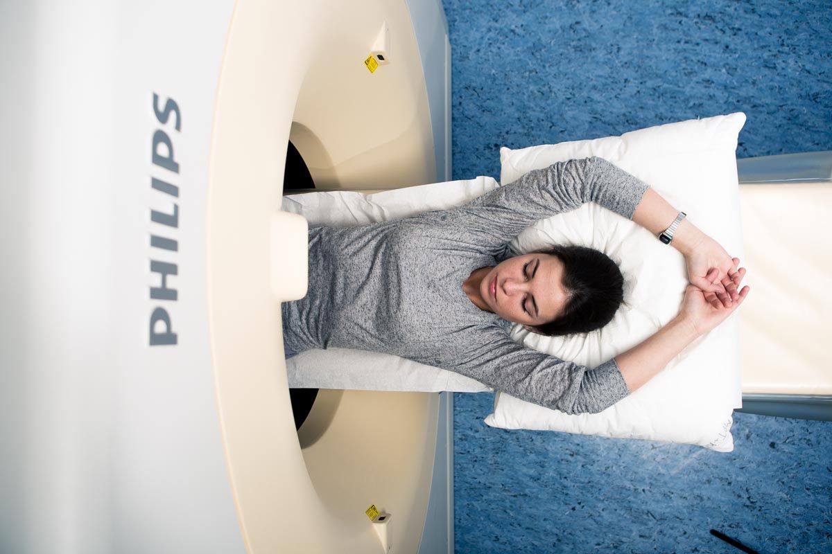
Computed tomography of the kidneys is performed at the European Medical Center using modern equipment that fully complies with European quality and safety standards. The Phillips iCT 256 multispiral CT scanner combines the ability to obtain excellent quality images with maximum patient comfort. Our MSCT device is equipped with the IDose software package, which significantly reduces radiation exposure during research.
One of the advantages of performing kidney CT at the European Medical Center in Moscow is the round-the-clock operation of the radiology department.
The department is headed by Professor Evgeny Isaakovich Libson (Israel). He is an honorary member of King's College (Great Britain), a member of the European and North American Radiological communities, and before joining the EMC, he headed the Radiation Diagnostics department of the Hadassah Medical Center, one of the largest in Israel.
The EMC uses modern research protocols that comply with the recommendations of the American and European radiological communities ESR, RSNA.
The impeccably polite and attentive EMC administrative staff does everything to make you feel comfortable and safe throughout your stay at the clinic.
Computed tomography of the kidneys with contrast
In some cases, a CT scan of the kidneys with intravenous contrast is necessary for better visualization of vascular structures and urinary tract (calyx-pelvis complex, ureters). At the same time, an iodine-containing contrast agent is used, which makes it possible to more clearly examine the small structures of organs, assess blood supply, urination, and other features of kidney functioning.It is only with contrast that it becomes possible to study lymph nodes, which is very important in the diagnosis and staging of kidney cancer.
The radiologist decides on the need for contrast based on the research objective.
Contraindications
Discuss with your doctor the possibility of performing a CT scan of the kidneys with contrast if you are pregnant, or if you
have:
- kidney failure;
- diabetes mellitus;
- diseases of the thyroid gland;
allergy to iodine and iodine-containing drugs.
In some cases, tests and medical training are required before the examination, but sometimes computed tomography has to be abandoned by choosing a different examination method.
Research progress
Before the examination, you will be asked to remove metal objects that fall into the scanning area. The X-ray technician will tell you how the procedure will take place and answer your questions. You will need to lie down on the CT scanner table. If necessary, to make it easier for you to fix a fixed position, the X-ray technician will offer you special pillows and rollers. Next, the X-ray technician will manage the study from the control room. You will hear it on the speakerphone. The table will enter the tomograph arch, after which the X-ray technician will take pictures of the required area. The study is conducted with a short-term respiratory arrest.
If necessary, after the first series of images, you will receive an intravenous contrast agent and continue the study.
The entire study will take no more than 15 minutes.
Results of computed tomography of kidneys
The images from the CT scanner are transmitted to professional workstations of radiologists. Most of the studies in our center are independently analyzed by two specialists, after which you are given a final conclusion. If necessary, the research data can be discussed within the framework of consultations of doctors (in this case, urologists and radiologists)
.
The time for the results to be ready depends on the complexity of the clinical situation. The doctor will tell you about the preliminary results immediately after the examination.
You can get the results in any form convenient for you.
Factors influencing the outcome of the study
The quality of images obtained with kidney CT may be affected by the patient's movement during the examination, the presence of surgical clips or catheters, and barium suspension in the gastrointestinal tract after radiography.
Computed tomography of kidneys for children
Both adults and children, including newborns, can undergo computed tomography of the kidneys at the EMC. To obtain clear images, the patient's immobility during the examination is of great importance. For active children, being at complete rest often becomes an impossible task, so the EMC has the opportunity to perform such studies under medication in the presence of an experienced anesthesiologist. In addition, one of the parents will be able to stay with the child during the study. It is only necessary to wear an apron that protects against X-rays.