Laparoscopic kidney resection
Radical nephrectomy, which was previously considered the "gold" standard of treatment for localized renal cell carcinoma, has now been replaced by laparoscopic renal resection (LDR). This is a low-traumatic organ-preserving operation aimed at excision of pathologically altered organ tissues. Thanks to the gentle technology, the functionality of the operated kidney is fully preserved. Laparoscopic partial nephrectomy is performed within the boundaries of a healthy parenchyma. The organ can continue to function fully, which is especially important if the second kidney is affected or missing. This is a modern, gentle method of fighting renal cell carcinoma.
Learn more about the methodology
Laparoscopic resection of the kidney is indicated for kidney tumors. Previously, the entire organ was removed during tumor processes. Radical intervention was considered the most effective way to treat malignant neoplasms. Since the kidneys are a well—supplied organ with blood, it was considered less risky to remove an organ with a tumor, leaving a person with one healthy kidney. Fortunately, medical technology is constantly evolving.
Improved diagnostic methods make it possible today to detect renal cell carcinoma at an early stage of development. This makes it possible to use a less radical approach to treatment — in most cases, only the tumor is removed, leaving the organ intact. The smaller the tumor, the higher the success of the operation. LDL has become the standard of treatment for exophytic neoplasms of small size (up to 4 cm).
Our clinic has significant experience in laparoscopic surgery, which allows the removal of tumors larger than 4 cm (even inconveniently located), without ischemia, in patients with early renal cell carcinoma. During the intervention, a laparoscope with powerful optics is used, which ensures the safety of the operation and allows performing all surgical procedures with precision.
Advantages of partial nephrectomy
- maximum preservation of healthy tissues;
- minimal risk of vascular damage and prevention of possible bleeding;
- absence of complications;
- the long-term result of the intervention is similar to radical nephrectomy;
- oncological safety of the patient;
- rapid rehabilitation.
What indications should I take
Partial nephrectomy is indicated if the lesion is localized in any segment of the organ. The indication for laparoscopic kidney resection is:
- renal cell carcinoma at stages T1 and T2;
- complicated renal cyst;
- traumatic injury to a part of an organ;
- benign tumor
The threshold size of a kidney tumor, after which resection is usually not performed, is a diameter of 5 cm. In addition to the size, the location of the neoplasm, proximity to blood vessels, and the cup-pelvis apparatus are important. Depending on this, the possibility of resection is discussed individually.
Contraindications
There are no absolute contraindications. The exception is somatically burdened patients who have difficulty undergoing surgery — cardiovascular, endocrine and other pathologies in the decompensation stage. But with the current level of medicine, such cases are extremely rare.
Why do I need surgery
Many nephrological pathologies respond well to conservative treatment, but in the case of a tumor process, the only way to get rid of the disease is surgery. Malignant neoplasm is a direct indication for surgical intervention. Only the removal of the altered tissues will stop the spread of the pathological process.
A characteristic difference between kidney cancer and malignant tumors of other organs is that the neoplasm remains clearly bounded from the healthy parenchyma by a pseudocapsule for a long time. Thanks to this capsule, the tumor does not grow into nearby tissues and organs for a long time, making the operation easier. Timely resection provides a high chance of a complete cure, without the need for radiation or chemotherapy.
Using a 3D modeling program, you can create a three-dimensional model of a kidney with a cancerous tumor, which clearly shows the vascular system, renal pelvis, and adjacent organs. This allows the surgeon to accurately plan the course of the operation, assess the risk of damage to important structures, the viability of the remaining part of the organ, and make a forecast for the further course of the disease.
Preparation, diagnostics
In addition to the standard diagnosis required for any surgical procedure, there are a number of studies that are required directly when planning laparoscopic resection. The list of studies is formed individually for the patient, taking into account the stage of the disease, the presence of concomitant pathologies. Preoperative examination involves careful preparation, assessment of the arteries, veins of the kidney, and individual characteristics of the vascular system. The pre-intervention examination algorithm includes:
- MSCT of the kidneys with contrast — allows you to get maximum information about the anatomy of the kidney and neoplasm. Based on the data obtained, the surgeon builds a 3D model to plan the course of the intervention.
- Nephroscintigraphy is aimed at assessing kidney function. It is not performed for small tumors if the condition of the organ does not cause the doctor any questions or doubts about the CT results. In other situations, the technique helps the doctor to determine the expediency of preserving the kidney, to understand how high the risk of acute renal failure is in the postoperative period. FVD (External respiration function) — this test allows you to assess the respiratory volume of the lungs, detect ventilation dysfunction and take timely measures to eliminate possible breathing complications during surgery. Ultrasound of the veins of the lower extremities — diagnosis of varicose veins, detection of blood clots, for the prevention of postoperative complications such as PE (blockage of the pulmonary artery or its branches by blood clots).
The list can be supplemented by other studies, such as ECHO—KG, Holter monitoring, EGDS, bicycle ergometry, etc. on the recommendation of an oncologist, therapist, cardiologist. Taking into account the expected blood loss, a reserve of donor plasma and erythrocyte mass is required.
Performing the operation
Laparoscopic kidney resection is performed under general anesthesia. During the intervention, the doctor mobilizes the vascular pedicle, applies a clamp to the artery, removes pathologically altered tissue within the boundaries of a healthy parenchyma (up to 1 cm around the neoplasm), and sutures the parenchymal defect. The blood flow of the kidney stops temporarily. A period of no more than 20 minutes is considered safe. In our clinic, resection usually takes no more than 10 minutes, so healthy kidney tissues are practically not injured. After resection, the organ functions unchanged.
If the tumor has slightly grown into the parenchymal tissue, resection can be performed without constricting the renal arteries. Excised tissues are sent for histological examination. Since the intervention is performed through 3 punctures of the abdominal wall, complex sutures are not needed.
Rehabilitation
The postoperative period is easy. The patient spends the first day after the intervention in the intensive care unit, under the supervision of medical staff. It is allowed to get up on the first day, and the patient is discharged on an outpatient basis for 2-3 days. During the first few days, some patients may have fever (up to 38°C). This does not indicate an infection, but is a natural reaction of the body to the operation. The stitches are removed before discharge, sterile bandages are applied to the wound, which the patient removes independently after 1-3 days.
Recovery after resection does not involve severe restrictions. However, within 3-4 weeks it is recommended to:
- limit physical activity to moderate (intense exercise, weight lifting, running, squats, etc., may cause bleeding);
- follow a gentle diet (exclude fried, smoked, salty, spicy foods, soda, alcohol), and a drinking regime.
Complete rehabilitation for about 2-3 months – when you can return to heavy physical activity. loads, as before the operation.
Get help
Specify your contacts and we will contact you to clarify the details.
Doctors

Grigorev Nikolay
Head of the Urological Clinic, Head of the Department of Urology at the EMC Medical School, Doctor of Medicine, Professor
-

Iskander Abdullin
President of the Association of Young Urologists of Russia (AMUR, Ph.D. of Medical Sciences
-

Ofer Yossefovitz
Professor, Doctor of Medicine
-
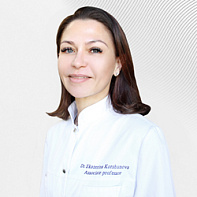
Ekaterina Korshunova
Ph.D. of Medical Sciences, Doctor of the highest category
-
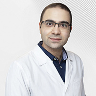
Vagan Barsegyan
Ph.D. of Medical Sciences
-
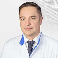
Aleksey Kovalenko
-

Gvozdev Mikhail
Doctor of Medicine, Professor, Honored Doctor of the Russian Federation, Doctor of the highest category
-
.jpg)
Tihonova Lyudmila
-
.jpg)
Gvasalia Badri
Professor, Doctor of Medicine
-
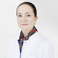
Sabirzyanova Zukhra
Ph.D. of Medical Sciences
-
Rubanov Valentin
Ph.D. of Medical Sciences
-
.jpg)
Vsevolod Matveev
President of the Russian Society of Oncourologists, expert of the European Association of Urologists (EAU) section "Prostate Cancer", Doctor of Medicine, Professor
-
Tedeev Rustam
Ph.D. of Medical Sciences
-

Kuznetskiy Yuriy
Doctor of Medicine, Doctor of the highest category, Professor
-

Dariy Evgeniy
Ph.D. of Medical Sciences, Doctor of the highest category
-
Grigorev Nikolay
Head of the Urological Clinic, Head of the Department of Urology at the EMC Medical School, Doctor of Medicine, Professor
- He is a recognized expert in endoscopic urology.
- He has performed more than 5,000 endoscopic and open surgeries on the organs of the genitourinary system
- Member of the Russian Society of Urology, the Russian Society for Endourology and New Technologies, the European Urological Association, the World Endourological Society
Total experience
34 years
Experience in EMC
since 2016


