Diagnostic endoscopy
Esophagogastroduodenoscopy
This is an endoscopic examination of the upper gastrointestinal tract, in which the diagnosis of the esophagus, stomach and duodenum is performed, and a biopsy is performed when visualizing pathological areas of the mucous membrane, including HP infection.
This procedure is associated with unpleasant and painful sensations. In Russia, gastroscopy is mainly performed without the use of anesthesia or with local anesthesia, in Europe and the USA - with the use of sedation, a type of anesthesia in which the patient is in a state of "drug-induced sleep."
EMC patients are relieved of discomfort during the study: short-term anesthesia (about 10-15 minutes), which is monitored by an anesthesiologist, provides a comfortable and calm state of the person, and the procedure is completely painless.
During the examination, an endoscopist can perform a biopsy of the mucous membrane with a test for Helicobacter pylori and remove polyps of the stomach and/or esophagus if they are found. The biopsy is sent to the EMC Pathology laboratory for histological examination, the results of which can be obtained within 3-5 business days.
Colonoscopy
It allows the most informative diagnosis of pathological changes and diseases of the colon in the early stages. Of course, this study is the prevention of colorectal cancer.
In EMC, colonoscopy is performed under medical sleep. To increase the effectiveness of the diagnosis, the chromoendoscopic method is used: the mucous membrane is stained with special dyes, which improves the ability to evaluate the visual picture and its interpretation, after which it is possible to select areas for targeted biopsy from pathologically altered areas. During the examination, the doctor can remove the identified tumors.
Bronchoscopy
The introduction of flexible endoscopes into the lumen of the trachea and bronchi makes it possible to diagnose diseases of these organs. This procedure is performed under medical sleep on an outpatient basis. During bronchoscopy, material is collected for all necessary laboratory tests. For certain diseases, a transbronchial lung biopsy or aspiration needle puncture biopsy of the lymph nodes can be performed.
Endoscopic ultrasonography of the tracheobronchial tree
Minimally invasive examination of the trachea and bronchi, which allows for the detection of neoplasms and simultaneous puncture to clarify the diagnosis. A bronchoscope equipped with an ultrasound sensor allows you to detect formations located around the bronchi, and simultaneously perform a puncture of these formations with a special needle under ultrasound control to clarify the diagnosis.The study is performed on an outpatient basis and does not require hospitalization. The patient is in a state of drug-induced sleep during the procedure. As a rule, endosonography of the tracheobronchial tree is performed immediately after traditional videobronchoscopy.
Endosonography (EndoUSI)
This method of examining the esophagus, stomach, duodenum, and pancreas, in which an echoendoscope (an ultrasound sensor in an endoscope) is inserted into the esophagus, further to the pathological focus. The method combines endoscopic and ultrasound examinations, which allows to obtain a clear picture of the structure of the walls of the organs of the gastrointestinal tract and anatomical structures, to identify and evaluate pathological neoplasms. During this diagnostic method, the doctor can perform a fine-needle puncture of the detected formation under ultrasound control.
Capsule endoscopy
A new diagnostic method for small intestine examination. An individual video capsule is swallowed by the patient on its own, passes through the gastrointestinal tract in 8-9 hours, takes pictures at a frequency of 2 frames per second, which are recorded in the memory of the receiving device through special sensors, and exits the body naturally. At the end of the study, the doctor evaluates the results using a special computer program.
Endoscopic retrograde cholangiopancreatography
Combined X-ray-endoscopic examination, in which an X-ray contrast agent is injected through an endoscope and the bile ducts and pancreatic duct are diagnosed under the supervision of an X-ray machine. The technique is used for diseases of the Vater's nipple, choledochus, and hepatic ducts. During the study, an extraction of stones from the common bile duct (choledochus) can be performed.
What is included in the cost of the survey
- preliminary consultation with an anesthesiologist and drug-induced sleep;
- disposable clothes and accessories for the procedure.
Preparation for endoscopic examination, documents for download
Get help
Specify your contacts and we will contact you to clarify the details.
Doctors
.jpg)
Danilov Dmirty
Ph.D. of Medical Sciences
-
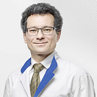
Butabaev Rustam
-
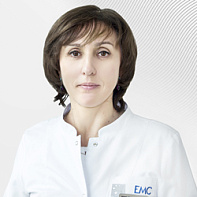
Elina Godzhello
Professor, Doctor of Medicine
-
.jpg)
Kim Denis
-
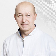
Filippov Mikhail
Ph.D. of Medical Sciences
-
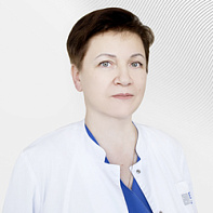
Logvinova Marina
-
Danilov Dmirty
Ph.D. of Medical Sciences
- More than 10 years of experience in emergency endoscopy: stopping gastrointestinal bleeding, removing foreign bodies from the gastrointestinal tract and bronchi
- Proficient in confocal laser endomicroscopy and capsule endoscopy
- He graduated from the Russian National Research Medical University named after N.I. Pirogov, completed his internship and residency in the specialty "Endoscopist"
Total experience
13 years
Experience in EMC
since 2017

