SPECT-CT
What is SPECT/CT
SPECT/CT (Single-photon emission computed tomography) is a highly sensitive diagnostic method that allows scanning the entire body, a limited area of the body, or a single organ.
OFFSET methods/CT scans are successfully used not only in oncology, but also in cardiology, endocrinology, traumatology and other fields.
For SPECT examination, a microscopic amount of a drug (for example, technetium) is injected into the patient. It actively accumulates in various organs and tissues of the body, is integrated into cellular metabolism, after which, immediately or after a certain time, depending on the type of study, the patient is scanned.
The image of metabolic disorders obtained by SPECT is superimposed on computed tomography (CT), which allows you to see a three-dimensional reconstruction of processes and anatomical changes. Thus, with a SPECT/CT scans can provide information about human disease at the molecular, cellular, and organ levels.
OFFER/CT scan at the European Medical Center
The cost of the study at the EMC Radionuclide Diagnostics Center includes:
- SPECT Study/CT scan on new equipment according to European protocols. All equipment is regularly checked and calibrated by our own engineer licensed by the equipment manufacturer.
- Additional SPECT/CT scan with contrast, delayed scanning*.
- High-quality radiopharmaceutical manufactured in our own laboratory and passed quality control in accordance with international standards.
- The report, written by 2 radiologists of the department, is consulted, if necessary, by Professor Libson (Israel).**
- The research disk.
- A printout with 3-dimensional reconstructions and key research frames.
- The opportunity to translate research into foreign languages.
- Attention and care at every stage of the study.
*The study is free of charge, and the decision to conduct additional research is made by the radiologist or the referring physician.
** The decision on the need for consultation is made by the radiologist of the department.
Indications for SPECT/CT
The Department of Radionuclide Diagnostics of the European Medical Center offers many types of SPECT studies/CT, including:
Sentinel lymph node scintigraphy
In oncology, sentinel lymph node scintigraphy is used to search for the sentinel (the first on the path of lymph outflow) lymph node in breast cancer, skin melanoma, and some other diseases. The purpose of this technique is to optimize and minimize surgical intervention in the absence of micrometastases in the sentinel lymph node.
You can read about sentinel lymph node biopsy in melanoma here. About sentinel lymph node biopsy in breast cancer – here.
Osteoscintigraphy
Skeletal bone scintigraphy is one of the most widespread studies in the field of radionuclide diagnostics. This study is used to diagnose the condition of bones for pathological processes. In addition, it helps to identify areas with pathological conditions such as fractures, inflammation, atrophy, and various tumors. The purpose of the bone scintigraphy study is to diagnose the causes of bone pain, monitor bone infections, identify bone metastases in cancer patients, identify traumatic processes, and so on.
Scintigraphy of the cardiovascular system
Radionuclide methods of studying the cardiovascular system make it possible to assess the functional state of the coronary arteries, the viability of the myocardium, and accurately determine indications for more invasive interventions such as angiography or heart surgery. In the EMC, studies of the cardiovascular system are conducted by a cardiologist-radiologist. The following techniques are available:
- Perfusion scintigraphy of the myocardium at rest
- Perfusion scintigraphy + exercise test on treadmill
- Perfusion scintigraphy + pharmacological stress test with (dipyridamole, adenosine, dobutamine)
- MUGA equilibrium ventriculography
Full list of SPECT studies/CT scans that you can undergo at a European Medical Center:
- Osteoscintigraphy
- Scintigraphy of the parathyroid glands
- Thyroid scintigraphy
- Sentinel lymph node scintigraphy
- Dynamic nephroscintigraphy of glomerular filtration
- Dynamic nephroscintigraphy of tubular secretion
- Static nephroscintigraphy
- Perfusion scintigraphy of the lungs
- Perfusion scintigraphy of the myocardium at rest
- Perfusion scintigraphy + exercise test on treadmill
- Perfusion scintigraphy + pharmacological stress test with (dipyridamole, adenosine, dobutamine)
- Salivary gland scintigraphy
Get help
Specify your contacts and we will contact you to clarify the details.
Doctors

Olga Penkova
-

Elina Moskalets
-
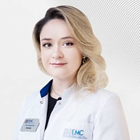
Olga Penkova
- Specialist in conducting and describing radionuclide studies
- Graduated from the Russian National Research Medical University named after N. I. Pirogov. Completed her residency at Russian Medical Academy of Postgraduate Education in the specialty: radiology.
- He holds regular annual internships at one of the leading centers in Europe: University of Belgrade (Belgrade, Serbia)
Total experience
7 years
Experience in EMC
since 2016


