Computed tomography (CT)
CT in EMC
- The most modern equipment that allows you to obtain the highest possible quality images with minimal radiation exposure is computer tomographs from Siemens and Philips, which have the latest radiation dose reduction systems (iDose7). CT is performed for both adults (including obese patients weighing up to 227 kg and patients with claustrophobia) and children, including newborns, during sleep or under anesthesia.
- The CT machine at the clinic in Orlovsky Lane is equipped with the Metal Artifact Suppression Program (iMAR) for orthopedic patients with metal implants in the body.
- Experienced anesthesiologists who will make the examination easy and comfortable.
About the study
Computed tomography (CT, MSCT) - a method of radiation diagnostics, which consists in a layer-by-layer examination of the human body using X-rays with the possibility of subsequent construction of multi-plane, including 3D images.
Today, this method has become one of the main ones in the diagnosis of most diseases and is widely used in both primary and clarifying diagnostics.One of the main advantages of computed tomography is the speed of the study and the possibility of simultaneous scanning of large anatomical areas.
Applications of CT diagnostics
A CT scan of the brain is a very fast and absolutely painless procedure that allows you to see changes in the brain that have occurred as a result of injury or infection. With the help of CT scans of the brain, it is possible to monitor the recovery of the organ after injury, and at the same time monitor whether complications develop.
CT of joints is performed according to the same principle as computed tomography of the brain, only it is not the head that is illuminated, but the joint.CT scans of joints are used to diagnose inflammatory processes, as well as the effects of injury.
In modern medical practice, CT techniques have almost completely replaced classical radiological ones.
- Many European clinics have almost completely abandoned X-ray imaging of the temporal bones in favor of CT.
- With recurrent diseases of the ENT organs (chronic sore throats, sinusitis, etc.).
- In children with suspected presence of foreign bodies in the lumens of the tracheobronchial tree.
- The "gold standard" for the examination of cancer patients is the assessment of the localization, shape, size, prevalence of tumor processes and the assessment of the dynamics of therapy.
- The primary method of examination of patients with severe traumatic brain injuries and any other traumatic injuries.
- It is indispensable for complex intra-articular fractures, confirmation or exclusion of epiphyseolysis (fractures peculiar only to children with the presence of growth zones in the bone structure).
- It is increasingly used to examine children with urinary tract pathology in studies with intravenous contrast.
Computed tomography (CT) with contrast is performed at the EMC.The contrast agent makes it possible to accurately determine the size, contours and location of the pathological formation. CT with contrast is used to examine almost all organs and systems of the body.
Methods of contrast agent management:
- intravenous,
- intravenous bolus,
- oral.
Contraindications to CT with contrast:
Iodine-containing substances are used for CT scans with contrast, so the indications for the study are always determined individually. It is important to exclude an allergic reaction to the contrast components. Also, before conducting the study, it is necessary to perform a biochemical blood test, since renal and hepatic insufficiency are contraindications for CT with contrast.
Types of research
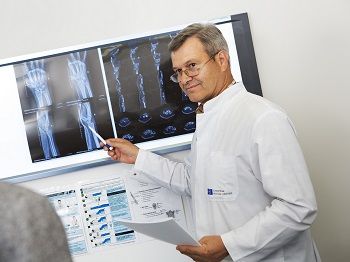 The following types of CT scans are performed at the European Medical Center:
- Computed tomography of the abdominal cavity
- Computed tomography of the brain
- Computed tomography of the sinuses
- Computed tomography of the spine
- Computed tomography of the kidneys
- Computed tomography of the bones of the skull/facial skeleton, orbits, jaw
- Computed tomography of the soft tissues of the neck/larynx
- Computed tomography of the chest and collarbone
- Computed tomography of the heart
- Computed tomography of pelvic bones
- Computed tomography of joints (CT of shoulder/shoulder joint, CT of forearm, CT of elbow joint, CT of wrist joint, CT of hand, CT of knee joint, CT of ankle joint, CT of hip joint, CT of hip, CT of shin, CT of foot, etc.)
- Computed tomography of the small pelvis
- CT colonography
- CT densitometry of the lumbar spine and femoral neck
- CT enterography
- Coronary calcium MSCT
- MSCT- coronary angiography
- MSCT angiography of the arteries of the upper/lower extremities
- MSCT angiography of the abdominal aorta and its branches
- MSCT angiography of the thoracic aorta
- MSCT angiography of the pulmonary arteries
- MSCT angiography of extra- and intracranial arteries
- Low-dose MSCT angiography of the pulmonary arteries
- Low-dose MSCT angiography of coronary arteries
Preparation for CT scan
The standard CT procedure (without contrast, without anesthesia) does not require special training from the patient and lasts no more than 40 minutes. Before performing the procedure, it is necessary to remove objects with metal elements that fall into the scanning area in order to avoid image quality degradation.
Important!24 hours before the study and within 48 hours after it, it is recommended to discontinue sugar-lowering drugs containing metformin.
CT scan of the abdominal cavity, pelvis and retroperitoneal space
The procedure is performed on an empty stomach or no earlier than 3-4 hours after the last meal. It is allowed to take medications (drink a small amount of water). Immediately before the examination, about 500-600 ml of water is drunk in the department.
CT scan of the urinary system
It is not recommended to empty the bladder half an hour or an hour before the examination.
CT of the heart and coronary arteries, CT angiography of any segment of the vascular bed
The procedure is performed on an empty stomach or no earlier than 3 hours after the last meal. On the day of the study, it is recommended to refrain from smoking, drinking coffee, tea and energy drinks.
CT enterography of the small intestine
The procedure is performed on an empty stomach, no earlier than 6 hours after the last meal. You should arrive at the department one hour before the scheduled examination time. Within an hour, you will need to drink 2 liters of Fortrans solution (1 packet per 1 liter of water) or mannitol solution (200 ml per 1.5 liters of water), 150-200 ml every 5 minutes. All medications are dispensed at the department.
CT scan of the colon (CT colonography)
Within 3 days before the study, it is necessary to follow a slag-free diet. Legumes, black bread, milk, carbonated drinks, vegetables, fruits, semi-finished products, and sweets are not recommended. Buckwheat, oats, lentils, rice, tea, fermented dairy products (if there is no intolerance), lean meat, fish, vegetable soups are allowed. The day before the study, it is necessary to take an Omnipack 350 solution (60 ml per 1 liter of water) in fractions during the day with meals (the drug can be obtained in advance at the Radiology Department). A light breakfast is allowed in the morning on the day of the study. It is necessary to arrive at the department 30 minutes before the start of the study.
Important!Don't forget to bring all the extracts, protocols, or recordings (discs) of previous studies. The more information the radiologist has before the examination, the clearer the task assigned to him. In addition, the previous results will allow us to assess the dynamics of the disease.
Prices for computed tomography with contrast can be specified inprice list of the EMC (Moscow). You can have a CT scan around the clock by appointment.
Doctors
.jpg)
.jpg)
.jpg)


.jpg)
.jpg)
- Actively participates in the work of scientific and research institutions
- He currently holds the position of Professor of Radiology at Hadassah Hospital. Member of the Education Committee of Hebrew University – Hadassah School of Medicine
- Awarded for outstanding contribution to Israeli Healthcare - Israeli Medical Association
