In The Neurosurgery Clinic of the European Medical Center under the guidance of a world-renowned neurosurgeon, MD, Professor, Corresponding member. RAN Alexey Leonidovich Krivoshapkin is undergoing surgical treatment of aneurysms and arteriovenous malformations of cerebral vessels of any complexity. The EMC Multifunctional Hospital on Shchepkina St., Moscow, has well-equipped neurosurgical operating rooms and an angiographic operating room with the latest generation Siemens Artis Zee Biplane system for performing both microsurgical and endovascular operations on brain vessels with the highest accuracy, which is very important in the treatment of brain aneurysms. Treatment at the clinic includes a full range of rehabilitation measures with the participation of rehabilitologists, physiotherapists, neuropsychologists, speech therapists and other specialists.
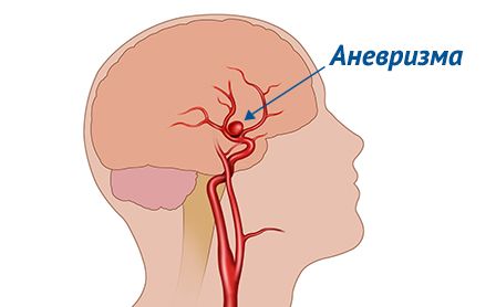 An aneurysm of the cerebral vessels is an expansion or protrusion of the cerebral artery wall.
Before rupture, aneurysms usually do not cause any symptoms and are diagnosed accidentally during examination for other reasons. Patients with a diagnosed aneurysm should be under medical supervision and undergo regular examinations to assess the progression of the disease.
Rupture of an aneurysm leads to intracranial hemorrhage. Most often, hemorrhage occurs in the subarachnoid space, the area between the brain and the arachnoid membrane.
Aneurysm rupture is a life–threatening condition requiring immediate medical intervention. When a brain aneurysm is diagnosed, treatment is performed using the most modern diagnostic and surgical equipment.
Aneurysm rupture
When an aneurysm ruptures, a sharp and very severe headache occurs. The patient may describe it as the most severe headache ever experienced.
In addition, rupture of a cerebral vascular aneurysm may be accompanied by:
-
loss of consciousness
-
blurred vision or diplopia (bifurcation of the visible image)
-
vomiting
-
nausea
-
photophobia
-
stiffness of occipital muscles
-
omitting the century
-
cramps
An unexploded aneurysm does not manifest itself in any way until, as it grows, compression of nearby nerves occurs. In this case, various symptoms may appear, including visual disturbances, eye pain, paralysis or numbness of the face.
The cause of the disease
The main cause of aneurysms is thinning of the vessel walls. Therefore, very often aneurysms appear at the site of the branching of the arteries, where the vessels become most vulnerable.
Usually, aneurysms form in areas of the cerebral arteries that run in the area of the base of the skull (the intracranial portion of the internal carotid artery, the middle and anterior cerebral arteries, the main artery and its branches).
Risk factors
Among the risk factors for aneurysms, there are congenital and acquired. Acquired risk factors are mainly related to lifestyle and concomitant diseases, such as:
-
atherosclerosis
-
arterial hypertension
-
smoking
-
drug use, especially cocaine
-
alcohol abuse
Congenital risk factors:
-
Hereditary connective tissue diseases such as Ehlers Danlos syndrome that weaken blood vessels
-
Polycystic kidney disease, a hereditary disorder that leads to the formation of multiple cysts in both kidneys and an increase in blood pressure
-
Pathological narrowing of the aortic lumen (coarctation of the aorta), the largest blood vessel in our body that carries blood from the heart throughout the body.
-
Arteriovenous malformation, a congenital malformation of the cerebral vessels, a disorderly intertwining of arteries and veins in the brain, which leads to disruption of normal blood flow.
-
Cases of aneurysm in the immediate family (parents, brothers, sisters)
Diagnosis of a brain aneurysm
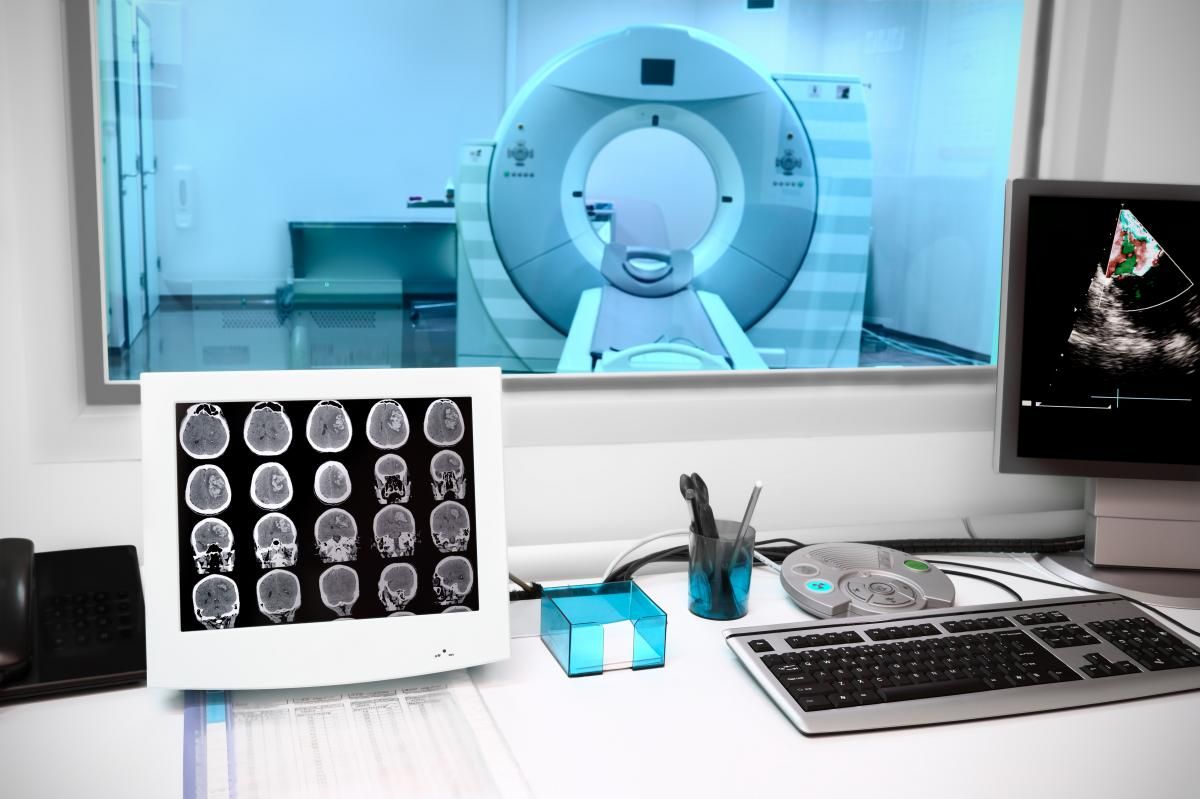 Aneurysm diagnosis is performed using modern instrumental and laboratory research methods.
A patient with a suspected ruptured aneurysm is first computed tomography, which allows to determine the presence of hemorrhage in the brain. At the same time, CT angiography can be performed with the introduction of a contrast agent to obtain an image of the cerebral vessels and detect the site of aneurysm rupture (CT angiography).
MRI is also used in the diagnosis of aneurysms. An MR scan allows you to obtain two-dimensional and three-dimensional images of the brain, and a detailed examination of the arteries (MR angiography) allows you to detect the site of aneurysm rupture.
Digital subtraction angiography of cerebral vessels – during this procedure, a catheter is inserted into the femoral artery, which is connected to the arteries in the brain. A contrast agent enters the vessels through a catheter, after which X-rays are performed. Angiography allows you to assess in detail the condition of the cerebral arteries and the location of the aneurysm rupture. This is an invasive procedure, and it is used in cases where other methods are not informative enough.
You can discuss with your doctor the need for screening examinations if your immediate family has had cases of ruptured aneurysms or if you have congenital risk factors for developing cerebral vascular aneurysms.
Aneurysm treatment
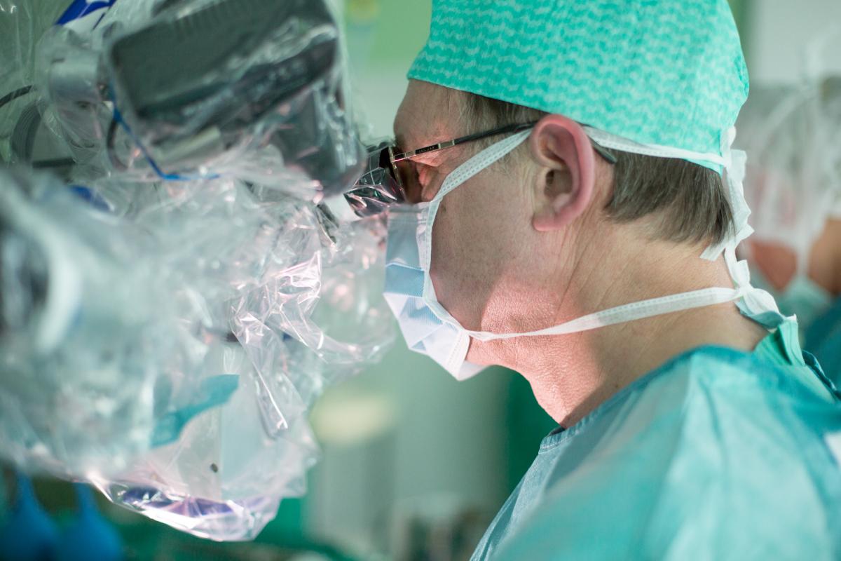 When a cerebral vascular aneurysm is diagnosed, treatment is performed in a neurosurgical clinic.
There are two main treatment options for aneurysms:
-
Clipping of an aneurysm is an open operation, the purpose of which is to turn off the aneurysm from the bloodstream by applying a special metal microplage (microclips) to the neck of the aneurysm at the point of its departure from the supporting artery.
-
Endovascular treatment is a minimally invasive operation in which microspirals are inserted into the aneurysm cavity to shut it off from the bloodstream.
The best option for surgical treatment of an aneurysm is determined by a neurosurgeon based on the size of the aneurysm, its location, the patient's state of health and other factors.


.webp)