Says a coloproctologist surgeon Ekaterina Borodina
 Rectocele" (rectocele: Latin. rectum – rectum; Greek. kele – protrusion, hernia, swelling) is a protrusion of the anterior wall of the rectum towards the vagina (anterior rectocele), and/or, extremely rarely, along the posterior semicircle of the rectum.
The vast majority of women suffer from rectocele. The weakness of the ligaments and muscles of the pelvic floor, which develops during life, and their damage during severe and complicated childbirth lead to a weakening of the rectovaginal septum — the wall between the intestine and the vagina. At the same time, it becomes thinner, and the posterior wall of the vagina, which is closely connected to the rectum, acquires pathological mobility. With an increase in intra-abdominal pressure (chronic constipation and severe straining during defecation), the anterior wall of the rectum bulges towards the thinned posterior wall of the vagina. Instead of straining to "work" on emptying the intestine, the septum protrudes into the vagina, the resulting pocket prevents normal bowel movements.
Rectocele is most often the tip of the iceberg of pelvic floor problems in women over 50 years of age and the causes of the so-called obstructive bowel movement syndrome. The failure of the pelvic floor muscles also leads to urinary incontinence during exertion (coughing, sneezing), and problems in the sexual sphere in women. That is why rectocele and omission of the female genital organs are diseases with a single mechanism of development and clinical picture, the adequate treatment of which requires the combined efforts of a coloproctologist and a urogynecologist.
Symptoms of rectocele
The main complaint with rectocele is constipation. Defecation is difficult, and there is a feeling of incomplete emptying of the rectum. As the disease develops, it becomes necessary to use manual assistance during stools and long stretches. Incomplete emptying of the rectum results in frequent, ineffective urge to defecate, and a need for two-stage bowel movements. A characteristic sign of the accumulation of feces in the "reservoir": the intestine "works" if you support or press your fingers on the back wall of the vagina or on the sides of the anus.
In the future, prolonged straining leads to injury to the mucous membrane of the anal canal and the occurrence of a number of concomitant proctological diseases (chronic hemorrhoids, anal fissure, rectal fistulas, chronic cryptitis, etc.).
Diagnosis of rectocele
 Typical complaints about difficult bowel movements, the need for manual assistance by pressing on the back the vaginal wall to release the rectum is a sufficient reason to assume that the patient has a rectocele. The main method of diagnosing a rectocele is a proctological examination performed on a gynecological chair in a supine position with legs bent at the knee joints and brought to the abdomen. When conducting a finger examination of the rectum and a vaginal examination, straining reveals a protrusion of the anterior wall of the rectum towards the vagina. An anoscopy/ rectoscopy/colonoscopy is mandatory for all diseases of the rectum and anal canal. At the same time, the condition of the rectum is determined and concomitant proctological diseases are detected. Transrectal ultrasound examination is performed to determine the condition of the pelvic floor muscles and the size of the rectocele.
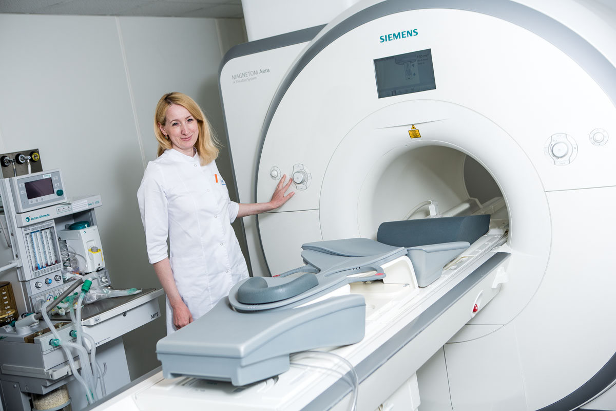
In specialized clinics dealing with pelvic floor problems, including the EMC Coloproctology Clinic, complex studies of the function of the act of defecation and pelvic anatomy are carried out using X-rays (defecography), MRI (MRI defecography), anal manometry. It is mandatory to examine the passage of radiopaque substance through the colon to exclude slow-transitory constipation.
Medical treatment
In all patients with rectocele, treatment should begin with conservative measures, consisting in the selection of a diet with the inclusion of high-fiber foods and a large amount of liquid in the diet. This is a diet with a high content of dietary fiber, medications with dietary fiber to soften and increase stool volume; eubiotics that stimulate the development of "beneficial" intestinal flora.
It has been proven that drinking liquids up to 1.5 - 2 liters per day increases the frequency of stools and reduces the need for laxatives in patients following a high-sugar diet. This therapy is aimed at normalizing the evacuation function of the colon, and it should be prescribed 1.5-2 months before surgery. In cases where intestinal function cannot be improved with the help of a diet, osmotic laxatives and prokinetics should be used — drugs that normalize the motor activity of the gastrointestinal tract.
In the initial stage, it is useful to perform a set of gymnastic exercises that strengthen the pelvic floor muscles (Kegel exercises).
Surgical treatment
If, despite all the measures taken to improve the emptying of the rectum, the symptoms of rectocele persist, the doctor is considering surgery to remove the protruding part of the rectum and strengthen the rectovaginal septum. This decision is made by a joint force of a gynecologist and a coloproctologist. Combined operations are possible. The choice of the surgical treatment method is carried out taking into account the patient's medical history and concomitant pathology. So, with pronounced pathological changes in the position of the pelvic organs or the presence of concomitant diseases, for example, cystocele, hemorrhoids, polyps or anal fissure, rectocele surgery is performed using combined access with simultaneous correction of concomitant diseases, since women suffering from rectocele often experience various damage to the anus, in particular, its anterior semicircle, inflammation of the sigmoid and rectum.
Before the advent of modern technologies and the latest materials, pelvic floor muscle plastic surgery was the main type of surgery. Modern technologies allow surgery to eliminate rectocele and rectal prolapse is laparoscopic and involves the installation of a mesh implant. Biologically inert implants are fixed on the ligaments of the small pelvis and reliably strengthen the rectovaginal septum, rectovaginal region and pelvic floor. The number of complications and relapses is minimized. A woman is allowed to get up the very next day after the operation and actively move for 2-3 days. The operation is low-traumatic and allows the treatment of older women who have suffered genital prolapse (vaginal prolapse and prolapse) and/or with a large number of concomitant diseases. In patients, normal pelvic floor muscle functions are restored and the bowel movement process returns to normal.
The method of mesh implant plastic surgery is not suitable for women who are planning pregnancy, as it becomes impossible to adequately stretch the rectovaginal region necessary for successful natural childbirth.
In cases where there are contraindications to surgery, urogynecologists decide on the appointment of special therapeutic exercises and recommend wearing a pessary – a rubber or plastic ring that prevents prolapse of the pelvic organs, including the uterus. The pessary supports a woman's internal genitals. It is installed in the vagina by a doctor and requires periodic replacement, as it causes the formation of pressure sores when worn for a long time.


.webp)


.webp)

.webp)
.webp)