Content
A patient with metastatic lung cancer underwent chemoradiotherapy and radiosurgery.
A 52-year-old man was diagnosed with adenocarcinoma of the upper lobe of the right lung with metastases to the lymph nodes of the lung root and mediastinum T3N2M, IV, EGFR, metastatic brain damage (solitary metastasis to the left parietal region). After the examination and consultation at the EMC, it was decided to give the patient chemoradiotherapy.
Radiation therapy was performed on the area of the affected lymph nodes and formations in the upper lobe of the right lung using the Varian Trilogy device using the RapidArc/IGRT technique, and after its completion, radiosurgery of the metastatic lesion in the left parietal region was performed using the Varian TrueBeam device using RapidArc/IGRT technology.
6 months after the treatment, no data were obtained for the presence of a tumor process in the right lung, lymph nodes and mediastinum, the available brain MRI data 3 months after the end of treatment indicated a decrease in the size of metastasis in the left parietal lobe, the presence of other pathological foci was not detected.
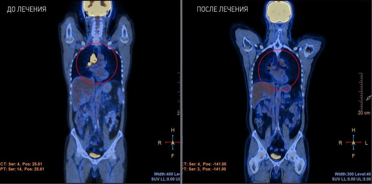
PET/CT data before treatment (06/09/2016)
A lesion of the right upper lobar bronchus (VDB) with metastases to the root and mediastinum has been confirmed. An MRI scan of the brain was performed for headaches, which revealed metastasis to the left parietal lobe of the brain.
PET/CT scan after chemoradiotherapy from 16.11.2016
The pattern of postradiation changes in the projection of the roots of both lungs, paramediastinally on both sides and in S6 of the right lung with increased metabolism. An intrathoracic (prevascular) lymph node with an increased metabolic rate. Effusion in the right pleural cavity and in the pericardial cavity. In comparison with the PET/CT study from 06/9/2016, the prevascular lymph node decreased in size, and the volume of formation in the root of the right lung was not reliably determined.
MRI of the brain from 11/16/16
There is a positive trend in the form of a decrease in the metastatic lesion of the left parietal lobe, no new foci have been identified.
PET/CT scan 6 months after treatment (dated 22.02.2017)
There was no evidence for the presence of foci of pathological metabolic activity characteristic of the 18F-FDG positive neoplastic process at the time of the study.
In comparison with the PET/CT study from 16.11.2016:
-
resolution of post-radiation changes in lung parenchyma;
-
absence of abnormal metabolism in the prevascular lymph node and reduction in size;
-
effusion in the pericardial cavity without dynamics;
-
No new foci of pathological metabolic activity of RFP have been identified.


.webp)