Get help
Specify your contacts and we will contact you to clarify the details.
Neoplasms of the mammary glands are the most common group of pathologies of the female reproductive system.80% of adult women are diagnosed with various types of tumors, and in the total structure of oncological diseases in Russia, the share of breast cancer is 20.9%. Modern diagnostic methods make it possible to identify and identify neoplasms in order to choose the optimal treatment tactics. One of the methods used to study breast tumors is biopsy, which involves taking a tissue sample for laboratory histological analysis. At the European Medical Center, all types of breast biopsies are performed using modern, high-precision equipment.
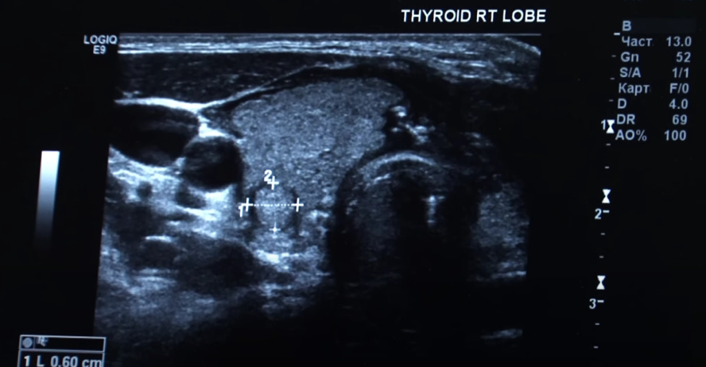
A breast biopsy is prescribed if a neoplasm is detected on ultrasound, MRI or mammography. The procedure is minimally invasive, it consists of taking a small fragment or part of the tumor under local anesthesia.
The material obtained during the biopsy makes it possible to determine whether the neoplasm of the mammary glands is benign or malignant, to establish the degree of malignancy and the HER2/neu receptor status (the level of tumor aggressiveness), sensitivity to estrogens (ER) and progestogens (PR). These data are necessary for choosing an adequate treatment strategy and (making) a prognosis.
Breast biopsy is possible at the EMC Mammology Center on the day of treatment, that is, almost immediately after mammography and ultrasound or MRI examinations.
The following types of breast biopsies are used in oncological practice: fine needle aspiration, vacuum, core biopsy. The choice of a specific type of study depends on the type of neoplasm, its location, and other factors.
The procedure is performed with a conventional syringe. Fluid is taken, for example, from a breast cyst for cytological examination. Puncture is performed only for fluid formations. Therefore, if we are not talking about cysts, a vacuum or core biopsy is required.
This is a percutaneous method of sampling for morphological examination with minimal invasion. The technique consists in inserting a tubular needle into the pathological formation using a mammotome vacuum device. Under the influence of vacuum, the material is attracted and separated into the needle cavity.
Traumatization of the surrounding gland tissues with such an intervention is minimal, and the patient will be able to quickly return to her usual lifestyle. The method has high information content and reliability — the accuracy of the study is 98%.
A biopsy is performed to make an accurate diagnosis. It can be assigned if:
detection of suspicious areas on a mammogram, MRI, or ultrasound scan;
the appearance of seals on the mammary gland;
the formation of crusts, peeling, redness, and the appearance of cavities on the skin of the breast;
nipple discharge not related to lactation.
Relative contraindications to breast biopsy are:
pregnancy and lactation;
acute infectious and inflammatory diseases;
Thrombocytopenia (low blood platelet count). In this case, preliminary monitoring and treatment of the patient in a hospital may be required.
The presence of breast implants is not a contraindication to a biopsy.
Before the biopsy, it is recommended to:
inform the doctor about chronic diseases and the therapy being received, allergic reactions, and the presence of a pacemaker in the case of an MR-guided biopsy.
stop taking blood-thinning medications at least one day before the procedure.
If additional training is required, the patient will be informed by the doctor at the consultation before the procedure.
Breast biopsy is performed on an outpatient basis under local anesthesia, which is selected individually. If the patient has previously been allergic to anesthetics, the procedure can be performed under general anesthesia.
Ultrasound, stereotactic (X-ray) equipment, or an MRI machine can be used for visual inspection.
The biopsy is performed in a supine position. The neoplasm is visualized using sonographic equipment, after which anesthesia is given and a sample is taken. The process takes no more than 15 minutes.
In this case, equipment similar to mammography equipment is used for visual inspection. The patient lies on her stomach on a movable stereotactic table with holes in the mammary glands. At the same time, the gland hangs down and is carefully fixed with a compression device. The device takes a series of images, which allows you to determine the coordinates of the formation and enter a tool to obtain samples. The preparation period for stereotactic biopsy lasts within 15 minutes.
Breast MRI is the most accurate and modern diagnostic method that allows you to accurately determine the location of the pathological zone without error. During the examination, the patient lies on her stomach on a movable tomograph table, the mammary glands hang down through the provided holes of the mammary gland coil. After the scan, the CT scanner builds an image of the tumor in several projections, and the doctor takes the biomaterial. The duration of the study is no more than 40 minutes.
The doctors of our clinic have the experience of performing the first breast biopsy in Russia under the control of MRI. To date, more than 400 MRI-controlled procedures have been performed at the EMC. For our doctors, this is a routine procedure that can be performed in all cases where there are indications for its implementation.
After a biopsy, a slight redness, bruise, or bruise may form at the puncture or incision site. Slight painful sensations are possible. To relieve discomfort, you can drink painkillers and make a compress with ice.
If there is severe swelling, bleeding, severe soreness, or fever, it is recommended to contact a clinic.
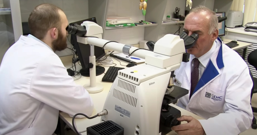
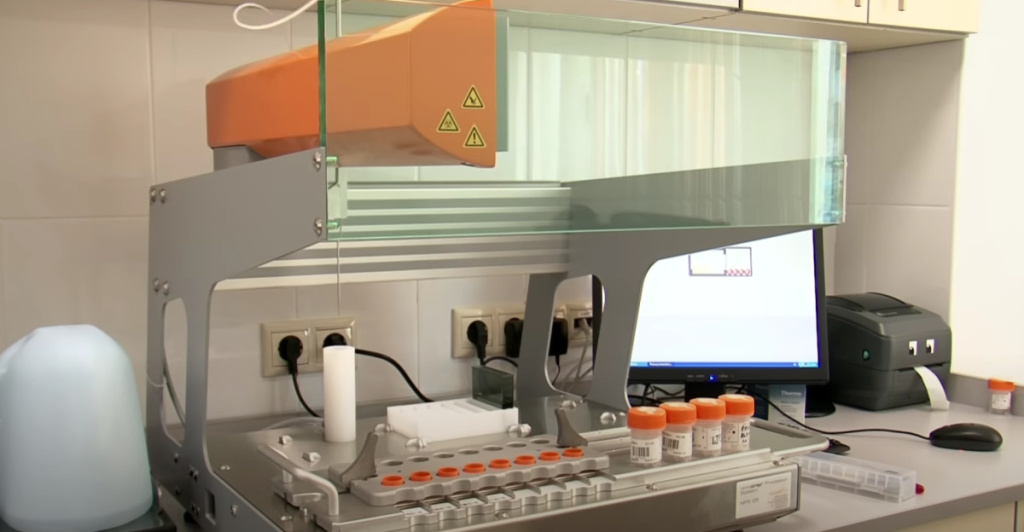
As a result of the biopsy, the number of tissue samples required for histological, immunohistochemical and genetic studies is obtained. The samples are sent for examination to EMC's own histological laboratory. The patient receives the result in 5-7 working days. If necessary, a second opinion from foreign experts can be obtained.
Based on the results of instrumental and laboratory studies, a multidisciplinary consultation of specialists is conducted The EMC Institute of Oncology to develop patient treatment tactics. The European Medical Center offers the most modern methods of diagnosis and treatment of oncological diseases: PET / CT, chemotherapy without hair loss, radiation therapy (including intraoperative), the most gentle surgery with subsequent breast reconstruction, if necessary.