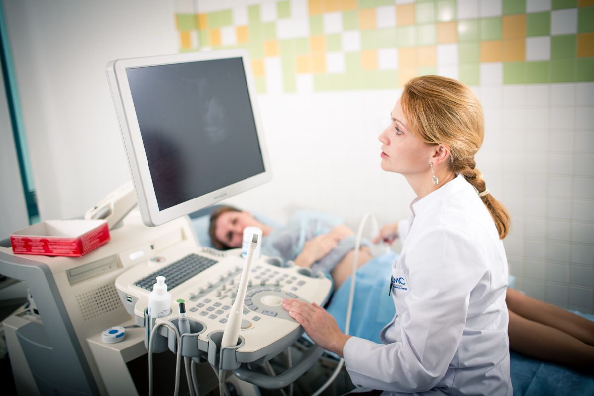
Expert ultrasound during pregnancy
According to the order of the Ministry of Health of the Russian Federation No. 572n dated November 12, 2012, screening ultrasound examination (ultrasound) of the fetus should be performed three times from 11 to 14, from 18 to 21 and from 30 to 34 weeks of pregnancy.
If necessary, additional ultrasound examination can be performed in any quantity and at any stage of pregnancy, taking into account the absolute safety of ultrasound for the fetus.
Ultrasound examination protocols at the European Medical Center are compiled depending on the timing of the study itself and based on the recommendations of the International Society of Ultrasound in Obstetrics and Gynecology (ISUOG), the Foundation for Fetal Medicine (FMF) and the Russian Association of Ultrasound Diagnostics in Perinatology and Gynecology. If necessary, all studies are accompanied by the use of 3D/4D technologies.At the end of the ultrasound, the expert doctor issues a study protocol with interpretation of the results.
Basic ultrasound examinations of the fetus during pregnancy
1. The first study to confirm the viability of the fetus.
This is an ultrasound examination, which is usually performed with a vaginal sensor at 6-10 weeks of pregnancy. The purpose of this study is to determine the number of embryos and confirm the normal development of the fetus inside the uterus.
This study is especially indicated for pregnant women with lower abdominal pain and/or spotting from the genital tract, as well as women who have had a history of undeveloped pregnancies, spontaneous miscarriages, or ectopic pregnancies.
2. Ultrasound examination for the period from 11 to 14 weeks.
Is usually performed transabdominally, but in some cases, if necessary, it can be performed transvaginally.
The purpose of the ultrasound at this time:
-
Set the exact gestation period. This is especially true for women who do not remember the date of their last menstruation, have an irregular menstrual cycle, or who became pregnant during lactation. By measuring the size of the fetus, we can accurately calculate the date of conception and the date of delivery.
-
Diagnosis of multiple pregnancies. Approximately 2% of independent pregnancies and 10% of pregnancies resulting from the use of assisted reproductive technologies are multiple pregnancies. Ultrasound allows you to determine whether both fetuses are developing normally, to assess the condition of the placenta (it can be the same for both babies, or each baby can have its own placenta), which is crucial for multiple pregnancies. In such cases, it is advisable to monitor pregnancy more closely.
-
Diagnosis of the main pathological conditions of the fetus. Some serious abnormalities and malformations may be visible during this period of pregnancy. However, it should be borne in mind that the main detailed ultrasound to exclude malformations is performed at a period of 20 weeks.
-
Diagnosis of early miscarriages. Unfortunately, 2% of women who come for a routine ultrasound at 11-14 weeks are diagnosed with an undeveloped pregnancy. Couples receive information about possible causes of non-developing pregnancies and additional examinations for planning a subsequent pregnancy.
-
Risk assessment of Down syndrome and other chromosomal abnormalities. Each woman is assessed individually for her risk. This indicator is calculated taking into account the age of the mother, the measurement of two hormones in the mother's blood and the results of ultrasound measurement of the thickness of the collar space, as well as measurements of nasal bones, assessment of blood flow in the fetal heart and venous duct, and assessment of fetal malformations. Parents will receive full advice on the significance of these risks and the need for more detailed studies - a non-invasive prenatal test or an invasive diagnosis - amniocentesis.
3. Ultrasound examination for a period of 20-24 weeks.
 The doctor examines each part of the fetus's body in detail, determines the position of the placenta, evaluates the amount of amniotic fluid and the degree of fetal growth. Special attention is paid to assessing the condition of the brain, face, spine, heart, stomach, intestines, kidneys and limbs.
4. Ultrasound examination to assess fetal development.
This ultrasound is usually performed between the 30th and 34th weeks of pregnancy.
The purpose of this ultrasound examination is:
-
Fetal head, abdomen, and femur measurements and fetal weight estimates.
-
Study of fetal movements.
-
Assessment of the position and condition of the placenta.
-
Measurement of the amount of amniotic fluid.
-
Assessment of blood flow in the placenta, fetal head, and uterine arteries using color Doppler mapping.
5. Ultrasound examination of the heart.
During ultrasound at 11-14 weeks, 20-24 weeks and 30-34 weeks, the doctor must assess the condition of the fetal heart and major vessels.
Expert detailed examination of the fetal heart is recommended:
-
women with a family history of congenital heart abnormalities, patients with diabetes mellitus, and those taking antiepileptic drugs;
-
in a fetus with suspected heart disease or in a fetus with an increase in the thickness of the fetal collar space, as well as in the case of other malformations in the fetus during routine ultrasound examinations.
The study is usually performed between 18 and 23 weeks of pregnancy, but can be performed as early as 13 weeks, if necessary.
6. Ultrasound examination of the cervix.
This is an ultrasound examination to measure the length of the cervix. It is recommended for women with a high risk of premature birth, multiple pregnancies; pregnant women with a history of premature birth, previous surgery on the uterus for malformations or surgery on the cervix.
This examination is usually performed for a period of 30-34 weeks, but if necessary, it can be performed starting at 16 weeks.
You can complete all studies during pregnancy in our maternity hospital.
Why the EMС
The first and only clinic in Russia, created in the image of the world's leading clinics


Get help
Specify your contacts and we will contact you to clarify the details.
Doctors

Vladimir Nosov
Ph.D. of Medical Sciences
-

Ron Schonman
Doctor of Medicine
-

Boris Sakhnovich
Doctor of Medicine
-

Dmitriy Subbotin
Ph.D. of Medical Sciences
-

Nenahov Filipp
Doctor of the highest category
-

Yashina Elena
-

Khachatryan Zarine
Ph.D. of Medical Sciences
-

Anoshin Alexey
-

Tyutyunnik Victor
Chief Doctor of the Perinatal Center, Doctor of Medicine, Professor
-

Sergunina Olga
-

Dikova Inna
Head of the Department of Pathology of Pregnancy
-

Trifanova Ekaterina
-

Saakyan Gayane
Doctor of the first category
-

Navrotsky Victor
Head of the Maternity Ward, Doctor of the highest category
-

Fateeva Irina
-

Alexeeva Inna
Head of the Obstetric Department, Doctor of the highest category
-

Polyakov Nikolay
-

Gorbunov Andrey
Doctor of the highest category, Ph.D. of Medical Sciences
-

Sukhobokova Elena
Head of the Neonatology Department with Intensive Care Unit and Pathology of Newborns, Doctor of the highest category
-

Sharova Marina
Head of the Mom School, Ph.D. of Medical Sciences, Doctor of the highest category
-
Vladimir Nosov
Ph.D. of Medical Sciences
- An expert oncogynecologist is a surgeon with more than 26 years of experience, including experience working in leading hospitals in the USA
- A leading Russian specialist in the field of robotic surgery in oncogynecology
- Graduated from the Moscow Medical Academy named after I.M. Sechenov
- Graduated from the Moscow Medical Academy named...
Total experience
27 years
Experience in EMC
since 2012