
Back pain is one of the most common complaints of patients. This is facilitated by a sedentary lifestyle, overweight, unreasonable physical activity, and sometimes a hereditary predisposition. The most accurate and safe method of diagnosing spinal problems is magnetic resonance imaging (MRI) .
Magnetic resonance imaging of the spine is a highly informative and safe diagnostic method. To put it simply, an MRI scanner is a large magnet, and the principle of obtaining images of internal organs is based on the use of a magnetic field.
MRI is widely used in neurology, oncology, and neurosurgery to diagnose and stage diseases of the spine and spinal cord, address the need for surgical intervention, and monitor its effectiveness.
Advantages of MRI of the spine over CT of the spine
Patients from other clinics often come to the EMC with a referral for a CT scan of the spine. However, computed tomography is not the method of choice for the primary diagnosis of spinal diseases. CT can be used for traumatic spinal injury as an adjunct to MR examination.
| MRI of the spine | CT scan of the spine |
| There is no radiation load at all | There is a radiation load due to the use of X-rays |
| Possibility of visualization of soft tissue structures of the spine (intervertebral discs, ligamentous apparatus of the spine, spinal cord) | Detailed visualization of the soft tissue structures of the spine is impossible |
| The possibility of a detailed assessment of soft tissue structures surrounding the spine (muscles and partially organs, for example, lungs at the thoracic level) | A detailed assessment of the soft tissue structures surrounding the spine is limited |
| The possibility of diagnosing vertebral fractures, including compression and stress fractures | Limited ability to visualize fractures, especially compression and stress fractures |
| The use of small volumes of contrast agents (no more than 15-20 ml) | Contrast agents are used in a volume of about 100 ml |
| Contrast media do not contain iodine, which makes them less allergenic | Contrast media contain iodine and may cause allergic reactions. |
Indications
- spinal injuries;
- dislocations and subluxations of the vertebrae;
- intervertebral disc protrusion and herniation;
- tumor, inflammatory and infectious processes in the spine and spinal cord;
- degenerative changes (osteochondrosis, etc.);
- vascular pathology of the spinal cord;
- deformities of the spinal column;
- narrowing of the spinal canal, compression of the spinal cord or nerve roots;
- congenital disorders of vertebral and spinal cord development.
Contraindications
An absolute contraindication to MRI of the spine is the presence of metal elements in the body, such as pacemakers, ferromagnetic implants, and vascular clips.
Be sure to consult with an EMC radiologist before performing an MRI of the spine if the pain syndrome does not allow you to remain stationary for a long time, you have heart failure, or if you are pregnant. All these are relative contraindications. Depending on the clinical situation, our specialist can offer you:
- postpone the study,
- to conduct a study using a different diagnostic method,
- to conduct the examination under anesthesia, accompanied by an experienced anesthesiologist,
- to perform anesthesia before the study.
How is the study going
Before the procedure, the assistant will ask you to fill out a questionnaire to identify possible contraindications. This is important for your safety. Next, you will be taken to the locker room and asked to remove all metal objects and jewelry. After that, you will need to lie down on the CT scanner table. The study can last up to 1 hour, so we make sure that you are as comfortable as possible. Pillows and cushions help to ensure a comfortable position. Since any movement you make during the examination can negatively affect the quality of the images obtained, the X-ray technician will fix your position using special devices. If contrast is intended to be used, a contrast agent will be injected into your vein through a catheter. You can signal any changes in your condition by squeezing a special warning bag and contacting an X-ray technician over the speakerphone.
MRI of the spine for children
In the European Medical Center, MRI is performed for both adults and children, including newborns. The study can be performed under medical sleep - experienced anesthesiologists work in the clinic.
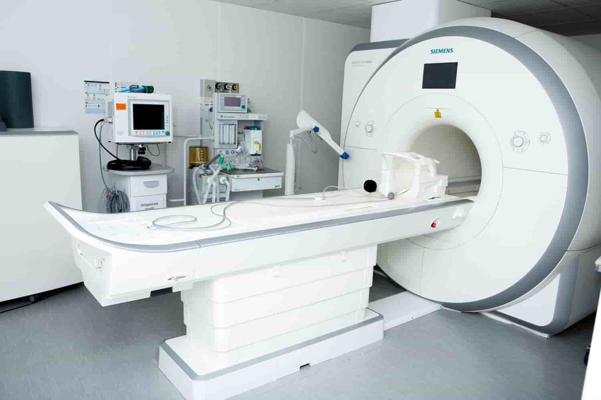
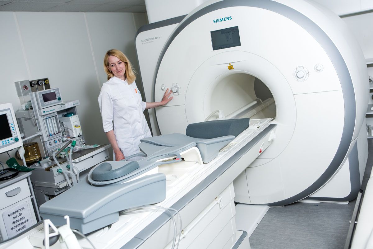
Equipment
A magnetic resonance imaging (MRI) scanner has been installed at the EMC clinic on Shchepkina Street The Siemens MAGNETOM Aera 1.5 T is a new–generation high-tech medical device with a wide tunnel, which makes the study comfortable for the patient.What can be determined by MRI of the spine
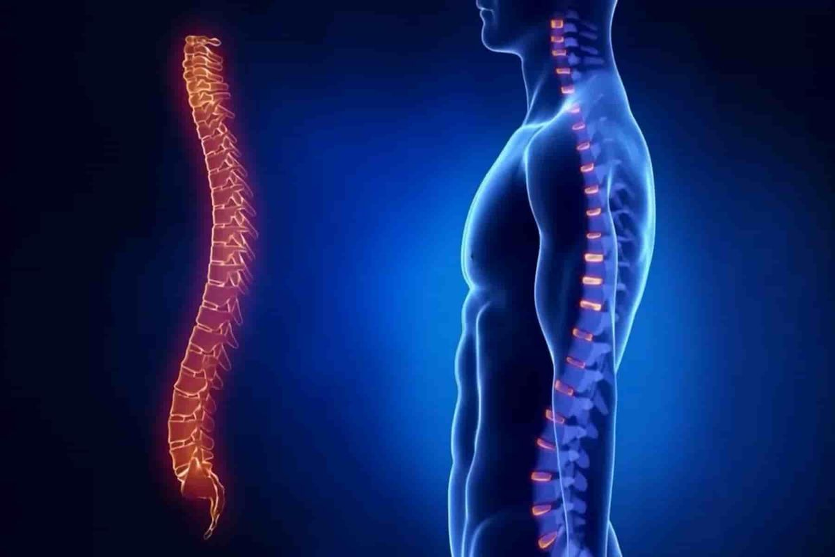
An MRI scan of the spine allows you to visualize not only the vertebrae, but also the intervertebral discs, nerves, blood vessels, joints, and spinal cord.
Using MRI of the spine, doctors can diagnose narrowing of the spinal canal and identify its cause, assess the condition of the intervertebral joints and amniotic tissues, and identify areas of the spinal cord with impaired blood supply at an early stage. In addition, MRI of the spine helps to simulate surgical procedures and evaluate their outcome.7 reasons to have an MRI scan of the spine at the European Medical Center in Moscow
- At the EMC, you can have an MRI scan at any convenient time, around the clock, on weekends and holidays.
- The Aera magnetic resonance imaging scanner from Siemens, installed at the EMC clinic on Shchepkina St., has a wide aperture ("tunnel") of 70 cm, the largest in this class of devices. This allows MRI scans to be performed on patients with large bodies (up to 150 kg) and makes the study more comfortable for patients with a fear of confined spaces.
- EMC doctors are specialists with extensive work experience who regularly upgrade their skills during internships abroad. The head of the EMC Radiation Diagnostics Department is Professor Evgeny Isaakovich Libson from Israel, formerly the head of the Radiation Diagnostics Department at Hadassah University Hospital (Hadassah).
- If necessary, the results of an MRI scan of the spine are evaluated by two radiologists (one of whom is a specialist in a specific anatomical area), after which the patients are given a final conclusion. Complex diagnostic cases are discussed in interdisciplinary consultations (with the participation of doctors of different specialties).
- EMC doctors use the PACS medical information system, in which the data of all studies are archived for at least 10 years. For patients, this is an opportunity not to worry about the safety of the images, and for their attending physicians, to view the images remotely and monitor the patient's condition over time during repeated visits.
- The EMC uses the most modern equipment. Maintenance of our CT scanners and software updates are carried out only by technical services authorized by the manufacturer.
- The impeccably polite EMC administrative staff does everything to make patients feel comfortable throughout their stay in the clinics.
Where can I get an MRI scan of the spine
MRI of the spine is performed in the clinics of the European Medical Center, located at the following addresses:
- St. Shchepkina, 35;
- Orlovsky Lane, 7.
Questions and answers
.webp)


.webp)


.webp)


.webp)

.webp)