Sometimes multiple IVF attempts with oocytes that are good from the point of view of embryology fail: for example, an implanted embryo does not take root and dies.
Why can this happen? The morphology of the oocyte division spindle is of great importance.
The structure of the egg, and what is the spindle of division?
The quality of the embryo and its development potential depend on the quality of the oocyte. The shape of the cell, the structure of the cytoplasm, the shell and the polar body (the cell separating from the oocyte during maturation) are usually evaluated. The egg itself may have an abnormal shape – oval or amorphous. The cytoplasm may have vacuoles (cavities inside the cell filled with cell juice), various inclusions, and an abnormal shape. The oocyte shell can be thick, thin, or carry any abnormalities: the polar body may be absent, fragmented, or located separately from the cell itself. All these anomalies affect the further development of the embryo to varying degrees.
There is another very important element in the oocyte – the spindle of division (Fig.1).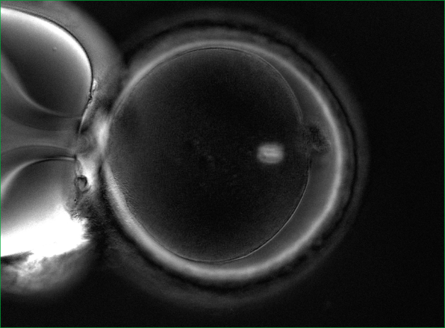
Who is shown the procedure for evaluating the division spindle?
1. For patients aged 40+. It is a well-known fact that with age, the percentage of oocytes with problems with chromosome separation increases. These problems lead to genetic mutations.
2. Patients with a large number of oocytes and poor embryo development (in particular, polycystic ovary syndrome).
3. Patients who have had multiple IVF attempts with good eggs from the point of view of embryology, but with embryos that do not take root after transplantation, or with early pregnancy losses.
How can the division spindle be evaluated?
It is impossible to see it with a conventional microscope, so no one evaluates it in routine practice. It is necessary to have a special polarizing installation for visualization. In world practice, only a few clinics have the necessary equipment in their arsenal and evaluate the spindle of division.
The importance of the quality of the division spindle in fertilization
The spindle of division can be either normal or abnormal. Normally, it should be clearly visualized and located close to the polar taurus. But there are various anomalies: vacuolization, the spindle is broken into fragments, poorly visualized, and may be completely absent (Fig. 2).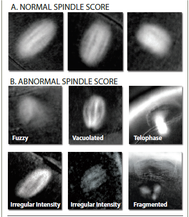 If the oocytes have a normal division spindle, the fertilization efficiency is 90%, and the embryo yield is 76%. In case of abnormal fertilization, the efficiency of fertilization is 72%, and the yield of embryos decreases to 31%. The probability of implantation of embryos that were obtained from oocytes with a normal spindle reaches 60%.
The size of the division spindle is also important. With a fission spindle from 90 to 120 microns2 , the chances of pregnancy are highest.
And one more important point: at the time of ICSI (injection of sperm into the cytoplasm), the embryologist must correctly position the oocyte. Usually, the marker is the polar body, under which the spindle of division should theoretically be located. In order not to damage the spindle, the oocyte is oriented for 6 or 12 hours, to maximize the removal of the spindle from the injection site. However, the spindle can be positioned in a cage in different ways (Fig. 3).
If the oocytes have a normal division spindle, the fertilization efficiency is 90%, and the embryo yield is 76%. In case of abnormal fertilization, the efficiency of fertilization is 72%, and the yield of embryos decreases to 31%. The probability of implantation of embryos that were obtained from oocytes with a normal spindle reaches 60%.
The size of the division spindle is also important. With a fission spindle from 90 to 120 microns2 , the chances of pregnancy are highest.
And one more important point: at the time of ICSI (injection of sperm into the cytoplasm), the embryologist must correctly position the oocyte. Usually, the marker is the polar body, under which the spindle of division should theoretically be located. In order not to damage the spindle, the oocyte is oriented for 6 or 12 hours, to maximize the removal of the spindle from the injection site. However, the spindle can be positioned in a cage in different ways (Fig. 3).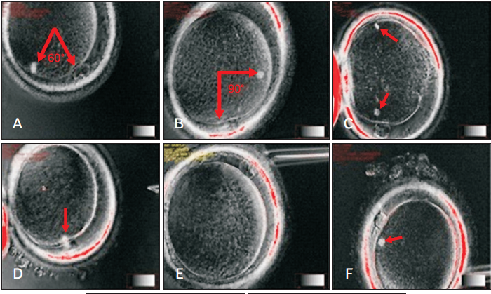 And since it is impossible to see the spindle during routine ICSI, there is a high probability of damage to it. The imaging system of the division spindle used in the EMC minimizes the risk of damage to it.
EMC capabilities in the evaluation of the division spindle
Specialists from the EMC Clinic for Reproductive and Prenatal Medicine conduct a detailed assessment of the spindle division. For this purpose, the European Medical Center has all the necessary equipment and experienced specialists who have worked and trained in leading clinics in Europe.
Who is shown the procedure for evaluating the division spindle?
1. For patients aged 40+. It is a well-known fact that with age, the percentage of oocytes with problems with chromosome separation increases. These problems lead to genetic mutations.
2. Patients with a large number of oocytes and poor embryo development (in particular, polycystic ovary syndrome).
3. Patients who have had multiple IVF attempts with good eggs from the point of view of embryology, but with embryos that do not take root after transplantation, or with early pregnancy losses.
How can the division spindle be evaluated?
It is impossible to see it with a conventional microscope, so no one evaluates it in routine practice. It is necessary to have a special polarizing installation for visualization. In world practice, only a few clinics have the necessary equipment in their arsenal and evaluate the spindle of division.
The importance of the quality of the division spindle in fertilization
The spindle of division can be either normal or abnormal. Normally, it should be clearly visualized and located close to the polar taurus. But there are various anomalies: vacuolization, the spindle is broken into fragments, poorly visualized, and may be completely absent (Fig. 2).
If the oocytes have a normal division spindle, the fertilization efficiency is 90%, and the embryo yield is 76%. In case of abnormal fertilization, the efficiency of fertilization is 72%, and the yield of embryos decreases to 31%. The probability of implantation of embryos that were obtained from oocytes with a normal spindle reaches 60%.
The size of the division spindle is also important. With a fission spindle from 90 to 120 microns2 , the chances of pregnancy are highest.
And one more important point: at the time of ICSI (injection of sperm into the cytoplasm), the embryologist must correctly position the oocyte. Usually, the marker is the polar body, under which the spindle of division should theoretically be located. In order not to damage the spindle, the oocyte is oriented for 6 or 12 hours, to maximize the removal of the spindle from the injection site. However, the spindle can be positioned in a cage in different ways (Fig. 3).And since it is impossible to see the spindle during routine ICSI, there is a high probability of damage to it. The imaging system of the division spindle used in the EMC minimizes the risk of damage to it.
EMC capabilities in the evaluation of the division spindle
Specialists from the EMC Clinic for Reproductive and Prenatal Medicine conduct a detailed assessment of the spindle division. For this purpose, the European Medical Center has all the necessary equipment and experienced specialists who have worked and trained in leading clinics in Europe.
And since it is impossible to see the spindle during routine ICSI, there is a high probability of damage to it. The imaging system of the division spindle used in the EMC minimizes the risk of damage to it.
Specialists from the EMC Clinic for Reproductive and Prenatal Medicine conduct a detailed assessment of the spindle division. For this purpose, the European Medical Center has all the necessary equipment and experienced specialists who have worked and trained in leading clinics in Europe.
Was this information helpful?
Questions and answers
How to lose weight correctly
I am 53 years old, my weigh is 116 kg. In 1988, my metabolism was probably, disturbed following the first childbirth. I gained weight, and since then I have been trying to fight with it periodically. I eat a lot of sweets however glucose is now normal and it always was. With age, my legs and back started to hurt me,
it is hard to “move” myself. I have undergone an examination in the district hospital, but nothing special was found. What tests and examinations I should bring to the doctor to get a diagnosis and treatment? Thank you!
...more Unfortunately, doctors not always can find the cause of excess weight. But this does not mean that there are ways to deal with it. Of course, it is not easy, but you definitely should try! At the first consultation it is desirable to have blood tests results, obtained no more than 6 months ago, total cholesterol,
glucose, glycated hemoglobin. Notes in the diary of blood pressure for 1-2 weeks are also important. Most likely, the doctor will arrange some additional examinations at visit, but everything is very individual and it's better to discuss all details with the doctor personally. You surely have to bring the results of previous all examinations with you.
...more 
Novikova Polina
08 September 2016
Question about ultrasound
I am 36 years old. Thyroid gland ultrasound: topography: position is regular, the right lobe is enlarged W-24 mm, t-22 mm, l- 51 mm, the volume is 12.90 cm3, left lobe enlarged, W -23 mm, t-23 mm, l-56 mm, volume is 14,90 cm3, isthmus thickness is 5 mm, the total volume is 27.09 cm3, alignments are even,
echostructure is inhomogenous, echogenicity is normal, focal masses are not seen, lymph nodes are not seen, conclusion; ultrasound signs of diffuse changes in the enlarged thyroid gland. TSH 2.10 mcIU/ml (normal range 0.30-4.00). The doctor prescribed Iodocomb 50/150 for 3 months. I have been taking the medicine for 2.5 months, but TG does not seem to diminish in size and I feel discomfort (it’s like a need to stretch my neck). Whether the treatment prescribed is correct? Are any additional tests needed? Should I have my thyroid gland diminished for those 2.5 months or it is too early to talk about it? Thank you!
...more It's hard to advice any treatment by correspondence. The cause of the thyroid gland enlargement is still unclear based on the results provided. The most common cause is iodine deficiency and Iodocomb treatment is appropriate in this case. Another cause is a chronic autoimmune thyroiditis which requires different
treatment. Endocrinologists at EMC will advise you and decide on the types of treatment and necessary doses.
...more 
Russ Irina
08 September 2016
Arthrosis
My left jaw started to hurt me after giving birth. The pain was accompanied by a crunch, clicks, discomfort when chewing. Arthrosis was diagnosed at local institution. What should I do?
The symptoms of pain and the crunch may occur following precipitating factors. It can be a trauma of the maxillofacial area, ENT-organs infections, sitting with mouth open for a long time when visiting the dentist, hypothermia, overload. Perhaps, pregnancy and childbirth were such a factor in your case. The treatment
goal in arthrosis is to reduce the load in temporomandibular joint. Treatment depends on the cause or the complex of causes that triggered the arthrosis. This may be due to the anatomical mismatch between the articular head size and the glenoid fossa. This also can be due to the long-term overload resulted from irregular bite and muscle imbalance. If increased tone of the masticatory muscles has led to increased abrasion of teeth, method of treatment with the restoration of the chewing surfaces and cutting edges of all teeth is optimal. But before that, splint therapy (mouth guards) is mandatory, which ensures the optimal position of the articular heads in articular fossae. Treatment is designed depending on the cause of changes in the joint. If the irregular bite is present pre-treatment by the orthodontist may be worthwhile.
...more
Problems with teeth
I have a huge problem with my teeth - all the teeth were treated, metalless ceramics were fixed in some places, a few teeth need to be removed. The treatment was carried out abroad, but the caries problem was beyond control, despite regular professional hygiene and specially selected tools. What range of dental
examinations, in addition to specialist’s consultation, I can count on in EMC Moscow?
...more Various dental diagnostic methods are available in our Dentistry Department: computed tomography of all teeth of both jaws, which gives a three-dimensional image and allows the doctor to more accurately diagnose; orthopantomography gives a flat image of all the teeth; the targeted x-ray tooth image. Tooth extraction
is performed surgically, mostly under local anesthesia. Diagnostic and treatment methods are determined individually by the attending doctor on consultation. Thank you for your message!
...more
How soon I may drive after LASIC?
When can I ride a car after LASIK?
Eyesight recovery is quite fast. Visual acuity is almost completely restored next morning, and most people can drive a car and get to work.

Elias Raid
08 September 2016


.webp)

.webp)