Sometimes multiple IVF attempts with oocytes that are good from the point of view of embryology fail: for example, an implanted embryo does not take root and dies.
Why can this happen? The morphology of the oocyte division spindle is of great importance.
The structure of the egg, and what is the spindle of division?
The quality of the embryo and its development potential depend on the quality of the oocyte. The shape of the cell, the structure of the cytoplasm, the shell and the polar body (the cell separating from the oocyte during maturation) are usually evaluated. The egg itself may have an abnormal shape – oval or amorphous. The cytoplasm may have vacuoles (cavities inside the cell filled with cell juice), various inclusions, and an abnormal shape. The oocyte shell can be thick, thin, or carry any abnormalities: the polar body may be absent, fragmented, or located separately from the cell itself. All these anomalies affect the further development of the embryo to varying degrees.
There is another very important element in the oocyte – the spindle of division (Fig.1).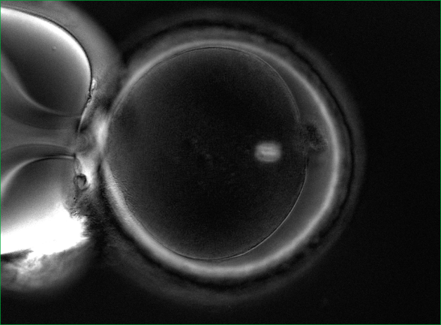
Who is shown the procedure for evaluating the division spindle?
1. For patients aged 40+. It is a well-known fact that with age, the percentage of oocytes with problems with chromosome separation increases. These problems lead to genetic mutations.
2. Patients with a large number of oocytes and poor embryo development (in particular, polycystic ovary syndrome).
3. Patients who have had multiple IVF attempts with good eggs from the point of view of embryology, but with embryos that do not take root after transplantation, or with early pregnancy losses.
How can the division spindle be evaluated?
It is impossible to see it with a conventional microscope, so no one evaluates it in routine practice. It is necessary to have a special polarizing installation for visualization. In world practice, only a few clinics have the necessary equipment in their arsenal and evaluate the spindle of division.
The importance of the quality of the division spindle in fertilization
The spindle of division can be either normal or abnormal. Normally, it should be clearly visualized and located close to the polar taurus. But there are various anomalies: vacuolization, the spindle is broken into fragments, poorly visualized, and may be completely absent (Fig. 2).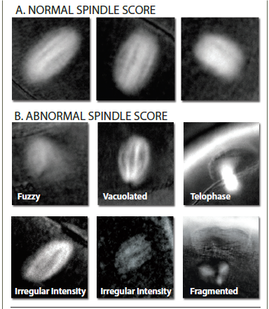 If the oocytes have a normal division spindle, the fertilization efficiency is 90%, and the embryo yield is 76%. In case of abnormal fertilization, the efficiency of fertilization is 72%, and the yield of embryos decreases to 31%. The probability of implantation of embryos that were obtained from oocytes with a normal spindle reaches 60%.
The size of the division spindle is also important. With a fission spindle from 90 to 120 microns2 , the chances of pregnancy are highest.
And one more important point: at the time of ICSI (injection of sperm into the cytoplasm), the embryologist must correctly position the oocyte. Usually, the marker is the polar body, under which the spindle of division should theoretically be located. In order not to damage the spindle, the oocyte is oriented for 6 or 12 hours, to maximize the removal of the spindle from the injection site. However, the spindle can be positioned in a cage in different ways (Fig. 3).
If the oocytes have a normal division spindle, the fertilization efficiency is 90%, and the embryo yield is 76%. In case of abnormal fertilization, the efficiency of fertilization is 72%, and the yield of embryos decreases to 31%. The probability of implantation of embryos that were obtained from oocytes with a normal spindle reaches 60%.
The size of the division spindle is also important. With a fission spindle from 90 to 120 microns2 , the chances of pregnancy are highest.
And one more important point: at the time of ICSI (injection of sperm into the cytoplasm), the embryologist must correctly position the oocyte. Usually, the marker is the polar body, under which the spindle of division should theoretically be located. In order not to damage the spindle, the oocyte is oriented for 6 or 12 hours, to maximize the removal of the spindle from the injection site. However, the spindle can be positioned in a cage in different ways (Fig. 3).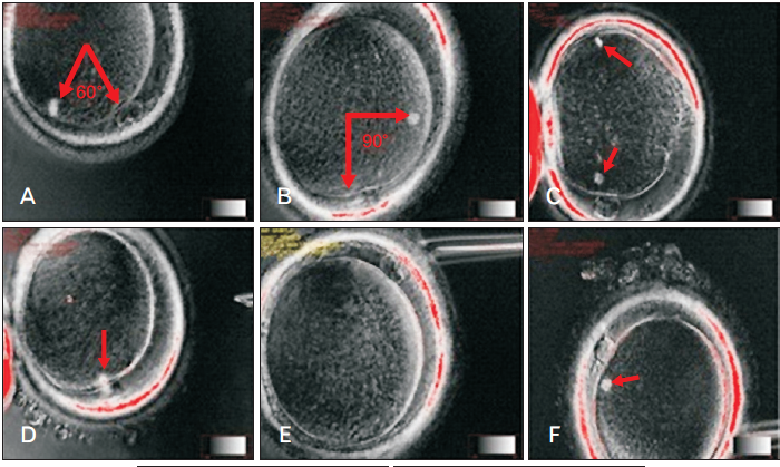 And since it is impossible to see the spindle during routine ICSI, there is a high probability of damage to it. The imaging system of the division spindle used in the EMC minimizes the risk of damage to it.
EMC capabilities in the evaluation of the division spindle
Specialists from the EMC Clinic for Reproductive and Prenatal Medicine conduct a detailed assessment of the spindle division. For this purpose, the European Medical Center has all the necessary equipment and experienced specialists who have worked and trained in leading clinics in Europe.
Who is shown the procedure for evaluating the division spindle?
1. For patients aged 40+. It is a well-known fact that with age, the percentage of oocytes with problems with chromosome separation increases. These problems lead to genetic mutations.
2. Patients with a large number of oocytes and poor embryo development (in particular, polycystic ovary syndrome).
3. Patients who have had multiple IVF attempts with good eggs from the point of view of embryology, but with embryos that do not take root after transplantation, or with early pregnancy losses.
How can the division spindle be evaluated?
It is impossible to see it with a conventional microscope, so no one evaluates it in routine practice. It is necessary to have a special polarizing installation for visualization. In world practice, only a few clinics have the necessary equipment in their arsenal and evaluate the spindle of division.
The importance of the quality of the division spindle in fertilization
The spindle of division can be either normal or abnormal. Normally, it should be clearly visualized and located close to the polar taurus. But there are various anomalies: vacuolization, the spindle is broken into fragments, poorly visualized, and may be completely absent (Fig. 2).
If the oocytes have a normal division spindle, the fertilization efficiency is 90%, and the embryo yield is 76%. In case of abnormal fertilization, the efficiency of fertilization is 72%, and the yield of embryos decreases to 31%. The probability of implantation of embryos that were obtained from oocytes with a normal spindle reaches 60%.
The size of the division spindle is also important. With a fission spindle from 90 to 120 microns2 , the chances of pregnancy are highest.
And one more important point: at the time of ICSI (injection of sperm into the cytoplasm), the embryologist must correctly position the oocyte. Usually, the marker is the polar body, under which the spindle of division should theoretically be located. In order not to damage the spindle, the oocyte is oriented for 6 or 12 hours, to maximize the removal of the spindle from the injection site. However, the spindle can be positioned in a cage in different ways (Fig. 3).And since it is impossible to see the spindle during routine ICSI, there is a high probability of damage to it. The imaging system of the division spindle used in the EMC minimizes the risk of damage to it.
EMC capabilities in the evaluation of the division spindle
Specialists from the EMC Clinic for Reproductive and Prenatal Medicine conduct a detailed assessment of the spindle division. For this purpose, the European Medical Center has all the necessary equipment and experienced specialists who have worked and trained in leading clinics in Europe.
And since it is impossible to see the spindle during routine ICSI, there is a high probability of damage to it. The imaging system of the division spindle used in the EMC minimizes the risk of damage to it.
Specialists from the EMC Clinic for Reproductive and Prenatal Medicine conduct a detailed assessment of the spindle division. For this purpose, the European Medical Center has all the necessary equipment and experienced specialists who have worked and trained in leading clinics in Europe.
Was this information helpful?
Questions and answers
How to stop dreaming?
Can you tell me if there are any medications that stop a person dreaming? I have been dreaming constantly for a long time, and the dreams are always vivid and emotionally charged. I wake up tired, not wanting to do anything, and I feel only lethargy and apathy. I live in another city.
We have all the tools for an accurate, comprehensive diagnosis, and medical treatment of such conditions, as well as psychosocial rehabilitation, which is critical for returning to an active lifestyle. The support program includes modern methods of diagnosis, regular professorial meetings, and comprehensive
psychological and neurocognitive support. Unfortunately, all the necessary diagnostic and therapeutic interventions can only be done with the patient being present at the Clinic. We are ready to welcome you to the EMC. Inpatient and outpatient psychiatry clinics operate within the multidisciplinary hospital, which creates the most comfortable conditions for patients.
...more 
Rivkina Natalya
09 November 2015
Baby teeth - treat or not?
Baby teeth do not need to be treated, because they will drop out in any case - is it true?
If the teeth are not cleaned properly white carious spots will appear underneath the soft plaque. This initial stage of decay is reversible: with proper hygiene the stain will gradually fade. The next stage is when the defect appears: the integrity of the enamel (the outer shell of the tooth ) is broken and a
cavity appears in the tooth . From this moment caries begins to develop more rapidly since the dentin (the hard tissue of the tooth located under the enamel) is less dense than enamel, and it becomes difficult to clean the teeth properly.
If microorganisms get into the cavity of the tooth where the nerve passes, this will cause pulpitis - inflammation of the pulp of the tooth (the pulp consists of blood vessels and nerves), which may be accompanied by pain, but in the milk teeth is usually asymptomatic.
Pain may occur later, when the nerve has died completely and infection occurs outside the tooth - in the bone. In addition to pain, pulpitis is sometimes accompanied by swelling of the gums, fistulas (channels between the foci of the infection and the gum) and loose teeth .
The pathological process - from the appearance of a white spot to the development of inflammation outside of the tooth root - can be fast and take a few months or last for years, depending on many factors.
To avoid problems with your child’s teeth , you should visit the paediatric dentist regularly. The first appointment with the dentist should be when the child is 1.5 years old. Following on from this there should be regular check-ups every 6 months, or more frequently as directed by the doctor.
...more
Diagnosed infertility - what to do?
I’m 27 years old, diagnosed infertility since 5 years ago, tubal factor and only one working ovary. Have gone through 3 tries of in vitro fertilization, one was stimulated, had no response, in all 3 cases I had only one oocyte. AMH 0,91. Do you think I have a chance for a pregnancy with my own oocyte? Sperm quality
is good.
...more
Yes, of course, you should fight for your oocytes. There is a variety of minimal stimulations: modified cycles, natural cycles etc. If these methods don’t work, so we will think about donor cells. But you must definitely give your ovaries a chance.
Severe cervical dysplasia
I have a transplanted kidney and I was recently found to have severe cervical dysplasia. The biopsy results are not yet back, but the physician says I must have my uterus and cervix removed. My question is: Can I have the operation in your clinic?
For severe cervical dysplasia, usually cervical conization is sufficient. If you have no plans for reproduction, or you already have children, then theoretically you can discuss having a laparoscopic removal of the uterus and cervix, but these decisions should not be made through correspondence. If you have a
referral for an operation in the city where you live, and have the opportunity to come to Moscow, come for a free consultation using "Second Opinion" promotion. If necessary, we can quickly provide operative treatment at a discount.
...more 
Vladimir Nosov
09 November 2015
Emergency's Working Hours
What are your hours?
We are open seven days a week, 24 hours a day, 365 days a year.


.webp)

.webp)