Breast cancer
The EMC Institute of Oncology provides comprehensive diagnosis and treatment of breast cancer according to modern American and European protocols (NCCN and ESMO) using modern methods of surgery, chemotherapy, targeted and hormonal therapy and radiation treatment. For our patients, this is a unique opportunity to gain access to the best breast cancer treatment methods without going abroad. The decision on treatment tactics for each patient is made at an interdisciplinary consultation (tumor board) with the participation of an oncologist, a chemotherapist, a surgeon, a mammologist, a radiologist, a histologist, a mammologist, if necessary, an oncopsychologist, as well as related specialists, are invited to the consultation. No more than 10-14 days pass from the moment of initial treatment to the operation, which is extremely important in the treatment of malignant diseases.
Breast cancer risk factors
 Breast cancer is the most common cancer in women in most developed countries of the world.
The incidence is particularly high in Europe and North America. In Russia, breast cancer also occupies the first place in the structure of oncological pathology in women. According to statistics, 55 thousand women get breast cancer in our country every year.
The presence of risk factors does not mean that a woman will necessarily get breast cancer. But knowing individual risk factors can help identify the disease at an early stage, when the disease is successfully treatable.
Breast cancer risk factors include:
-
Age: the risk of breast cancer increases when reaching the age of 45-50 years
-
Weight: excess weight is a risk factor for postmenopausal cancer, however, an increased body mass index coefficient is associated with a reduced risk of premenopausal breast cancer.
-
Elevated estrogen levels
-
High density of breast tissue
-
Hormone replacement therapy in menopause (prolonged, more than 5 years, taking combined estrogen-progestin drugs by women aged 50-79 years)
-
Reproductive factors: early onset of menstruation and late menopause, late childbirth and their absence, lack of breastfeeding
-
Heredity is one of the main risk factors. About 10% of all breast cancer cases are associated with inheritance of mutations in the BRCA 1 and BRCA2 genes. In women with a mutation of these genes, the risk of breast cancer during their lifetime ranges from 50 to 85%.
Lifestyle and nutrition features are also associated with increased risk, in particular:
-
Reduced physical activity
-
Smoking
-
Alcohol consumption
-
Eating red meat more than 5 times a week. According to research, this increases the risk of developing hormone-receptor positive breast cancer in premenopausal women.
According to some reports, a diet dominated by vegetables and fruits, as well as foods containing soy, may slightly reduce the risk of breast cancer. The role of antioxidants in reducing morbidity has not been proven. The effect of caffeine on morbidity has also not been scientifically confirmed.
Environmental factors:
-
Radiation therapy on the chest area in the past (for example, in the treatment of Hodgkin's lymphoma)
-
Night shift work, according to some reports, is a risk factor for breast cancer, which may be due to a lack of the hormone melatonin, which is produced only at night.
Breast cancer symptoms
In countries with a developed screening system, breast cancer is detected in most patients as a result of mammography. But in 30% of cases, a palpable tumor is detected between regular mammographic examinations, such cases have even received their name – "interval cancers" of the breast.
The "classic" signs of a breast tumor are a solid, motionless single lump with uneven edges. However, these signs do not allow us to judge the nature of education. Symptoms of locally advanced cancer may include:
-
enlargement of lymph nodes in the axillary area
-
thickening of the skin, the effect of "orange peel".
Warning signs also include visual changes to the nipple, such as its retraction or discharge from the nipple.
How to protect yourself from breast cancer?
Breast cancer screening
Unfortunately, the above symptoms are often a sign of a common process. Therefore, regular screening is important for early diagnosis of cancer. This is a study that detects the disease before its clinical manifestations.
Breast cancer screening begins at the age of 40-45 years and is carried out annually or every two years (international expert organizations give various recommendations). Therefore, in each individual case, it is better to determine the beginning of research and their frequency with a doctor.
The main research method accepted as a diagnostic standard is digital mammography.
Screening for women at risk begins earlier, usually 5 years before the age when the disease was detected in the next of kin. Our clinic has developed special screening programs for women with a genetic predisposition. According to international recommendations, they include magnetic resonance imaging with contrast imaging alternating with ultrasound of the mammary glands every 6 months.We also carry out molecular genetic diagnostics – BRCA 1 and 2 gene mutation test.
Breast cancer diagnosis
 Breast cancer diagnosis includes:
-
Clinical diagnosis: physical examination when a woman goes to the doctor;
-
Instrumental diagnostics: Breast ultrasound, mammography, MRI of the mammary glands.
Clinical examination is important, but its role is insignificant for the early diagnosis of breast cancer, since palpation does not detect small tumors or deeply located neoplasms.
Ultrasound of the mammary glands is performed for women at any age. Ultrasound is an "operator-dependent" method, so it is important that the study is performed by a qualified specialist using modern equipment. Ultrasound is only an additional research method in the diagnosis of breast cancer.
Digital mammography is the main method of breast cancer diagnosis. EMC performs digital mammography with tomosynthesis, a layered image of breast tissue.It has been proven that this technique increases the effectiveness of breast cancer diagnosis by 20% compared to conventional mammography. Digital mammography has age restrictions and cannot be used for diagnosis in women under 40 years of age.
An important pathognomonic, i.e. characteristic feature of breast cancer is the accumulation of microcalcinates visible in the image. Even those cases when there is no tumor node as such on the mammographic image, but only an accumulation of calcifications in a limited area, are indications for a therapeutic and diagnostic procedure - sectoral breast resection, since the tumor may be X–ray negative and a malignant tumor may be hidden among these calcifications - cancer in situ (noninvasive cancer).
Breast MRI with contrast is a highly informative method for breast diagnosis. The method is used in women with high breast density in cases of suspected breast cancer based on mammography results, as screening in young women with a genetic predisposition to breast cancer and in other complex diagnostic cases.
The final method of breast cancer diagnosis is a breast tumor biopsy.
A fine needle puncture biopsy (when a tumor is punctured with a thin needle of a medical syringe and the resulting cell material is sent for cytological examination) is not always sufficient to make a diagnosis. For a full-fledged diagnosis and selection of the correct treatment tactics, it is necessary to conduct a CORE biopsy, when tumor tissue is taken under local anesthesia of the skin of the gland for subsequent histological examination.
CORE biopsy is performed on an outpatient basis and allows us to determine not only the histological structure of the tumor, but also to conduct an immunohistochemical study to determine the most important characteristics of the tumor: the presence or absence of hormonal receptors, the HER 2/neu receptor, as well as the Ki-67 marker, the proliferation index, or the growth rate of tumor cells.
EMC Histological Laboratory has the highest ratings of the independent quality control organization for immunohistochemical research NordiQC in the field of breast cancer diagnosis. If necessary and at the request of the patient, the diagnosis is verified (histological preparations are reviewed) in leading European and American cancer clinics.
By contacting EMC Institute of Oncology, you can be sure that you will receive all the necessary diagnostic and therapeutic procedures according to modern international standards. If pathological areas are found and a biopsy is necessary, it is performed on the day of the first treatment, that is, immediately after the mammography. This approach not only saves you time, but also reduces unpleasant waiting and anxiety before the procedure.
Breast cancer staging
After diagnosis, it is necessary to determine the extent of the tumor process, since cancer has a tendency to metastasis. Even a small tumor can give distant metastases to other organs. Treatment approaches and prognosis of the disease depend on the stage.
The standard of examination is determined by the most frequent sites of breast cancer metastasis. As a rule, these are:
-
regional lymph nodes (axillary, supra- and subclavian intramammary lymph nodes),
-
lungs,
-
the second mammary gland,
-
liver,
-
bones.
Breast cancer screening may include:
-
CT scans of three areas: CT scan of the chest, CT of the abdominal cavity and CT of the pelvic organs;
-
All these studies can be replaced with one - PET/CT scan of the whole body.
According to statistics, PET/CT scans for breast cancer in at least 30% of cases change the initial treatment strategy, allowing you to choose personalized and effective treatment for each patient.
At EMC, PET/CT scans are performed by qualified radiologists using state-of-the-art equipment using high-quality radiopharmaceuticals from our own production and in the shortest possible time. Since 2016, patients who are registered or have permanent registration in Moscow can undergo PET/CT for newly diagnosed breast cancer for free under the compulsory medical insurance policy if there is a referral from an oncologist.
If a patient complains of dizziness, sudden unreasonable headaches, and a feeling of double vision, an MRI of the brain is performed after exclusion of high blood pressure and orthostatic collapse.
Assessment of axillary lymph node damage in breast cancer
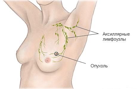 One of the main sites of breast cancer metastasis is the axillary lymph nodes located in the axillary region. The involvement of lymph nodes is one of the most important factors for determining the long-term prognosis of breast cancer and the choice of treatment tactics.
-
If tumor cells are detected in the lymph nodes, then there is a high probability of further spread of the tumor to various organs and tissues, therefore, in this case, adjuvant (postoperative) systemic chemotherapy is recommended for most women.
-
In the absence of lymph node involvement, systemic therapy is prescribed less frequently, especially with small tumor sizes, as well as with other prognostically favorable factors (for example, with estrogen receptor-positive tumors).
Meanwhile, adjuvant hormone therapy is given to all women with estrogen receptor-positive tumors, even in the absence of lymph node damage. Hormone therapy has fewer side effects compared to chemotherapy and significantly reduces the risk of cancer recurrence in the future.
Examination of axillary (axillary) lymph nodes
Examination of axillary lymph nodes is performed by visual examination, palpation of lymph nodes by an oncologist, ultrasound and biopsy. If there is a proven lesion of the lymph nodes, they are removed during surgery – axillary lymph dissection.
In patients with an early stage of the disease, if there are no signs of lymph node damage, a sentinel lymph node biopsy is performed. In this case, the first lymph node in the path of lymph outflow from the tumor (the so-called "sentinel") is removed, and its histological examination is performed. EMC is one of the few clinics in Russia where sentinel lymph node biopsy is performed for breast cancer.
If no metastases are found in the sentinel lymph node, the risk of damage to the remaining lymph nodes is minimal. In this case, diagnosis of the remaining lymph nodes or their removal is not required.
Thus, the procedure allows you to obtain important information for the staging of the disease, and at the same time avoid serious complications, in particular, lymphatic edema of the arm – lymphedema, which often occurs as a result of extended axillary lymph dissection.
Stages of breast cancer
Noninvasive breast cancer
The earliest stage of breast cancer is called "in situ" cancer. The term "in situ" means "in situ".
Ductal carcinoma "in situ" (noninvasive ductal cancer) is a noninvasive malignant neoplasm that develops in the mucous membrane of the ducts of the mammary glands.
The primary treatment of ductal carcinoma "in situ" is surgical removal of the affected breast tissue. The amount of tissue to be removed depends on the size of the affected area and breast, the degree of malignancy of the tumor, and the general condition of the patient. Most women undergo organ–preserving treatment – removal of a small area of the breast - lumpectomy followed by radiation therapy. In older patients with a small tumor and a low degree of malignancy, only surgical treatment can be performed, without radiation. In case of extensive ductal carcinoma, a mastectomy may be recommended, during which a sentinel lymph node biopsy is usually performed.
Patients with ductal carcinoma "in situ" do not receive chemotherapy. Hormone therapy is prescribed to women with estrogen receptor-positive tumors. The main hormone therapy drugs for premenopausal women are antiestrogens. Aromatase inhibitors are prescribed for postmenopausal women.
Lobular carcinoma "in situ" (lobular cancer) is not an oncological disease in the literal sense of the word and is rather considered as a precancerous condition in which pathological cells are located in the lobules of the breast.
Women who have been diagnosed with lobular cancer in situ should consult a specialist about receiving treatment aimed at reducing the risk of developing invasive cancer. Regular health monitoring, annual mammography, MRI and periodic breast examinations are required.
Stages of invasive breast cancer
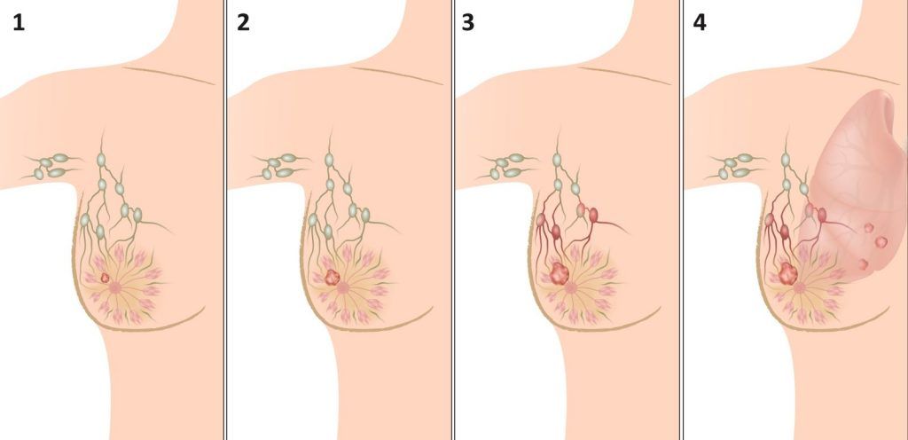 Breast cancer of stages 1 and 2 is called early stage cancer, or localized cancer.
Stage 1 breast cancer is a tumor of less than 2 cm, without damage to the lymph nodes.
Stage 2 breast cancer – the tumor has spread to the axillary lymph nodes and/or the tumor size is more than 2 but less than 5 cm.
Stage 3 breast cancer - it is also called locally advanced cancer. It is characterized by a large tumor (more than 5 cm in diameter), extensive metastasis to the axillary (axillary) lymph nodes (when more than 10 lymph nodes are affected), involvement of intramammary, supra- and subclavian lymph nodes.
In stage 3 cancer, the tumor can grow into the skin and into the muscle tissues located under the breast.
Inflammatory breast cancer is a fast–growing type of tumor that causes redness and inflammation of the breast. It belongs to stage 3 cancer, even with a small tumor size and the absence of lymph node damage.
Stage 4 breast cancer – the tumor metastasizes to distant organs – bones, lungs, liver, etc. In this case, the size of the initial tumor and the degree of damage to the lymph nodes can be any.
Factors affecting breast cancer treatment
To date, breast cancer treatment is personalized, it depends not only on the stage of the disease, but also on the molecular biological subtype of cancer - the totality of the molecular characteristics of the tumor. An immunohistochemical study allows us to answer the question of whether estrogen and progesterone receptors, the HER2/neu receptor, and the proliferative index of the tumor Ki-67 are present in the tumor.
Generally speaking, tumors that express sex hormone receptors and do not express HER2/neu receptors have a more favorable clinical course. Such tumors, as a rule, respond well to hormone therapy and are associated with a better prognosis of the disease.
If the HER2/neu receptor is expressed in the tumor, the woman is prescribed targeted therapy with herceptin for one year.
Triple negative breast cancer is a tumor in which there are no receptors for estrogens, progesterone and HER2/neu. As a rule, these are tumors with a high Ki-67, i.e. with a high proliferation index, which means that the cells divide very quickly. This tumor is considered the most unfavorable and has an extremely high metastatic potential. But, on the other hand, such tumors are most sensitive to chemotherapy.
The molecular biological subtype of the tumor also suggests the most common sites of cancer metastasis. Triple negative tumors most often metastasize to the central nervous system, for example, to the brain. Therefore, when a woman with triple negative breast cancer complains of unsteady gait and dizziness, she should definitely have an MRI scan of the brain. Hormone receptor-positive breast tumors are more likely to metastasize to the bone system.
Modern genetic analysis Oncotype DX (Oncotype), USA, is available to patients of the EMC Institute of Oncology. Based on the analysis of the expression of a group of genes, the test allows us to assess the individual risk of cancer recurrence in a patient with early-stage breast cancer and decide whether to undergo chemotherapy.
Treatment of stage 1 and 2 breast cancer

The treatment of patients with stage 1 and stage 2 breast cancer is almost identical with minor differences. There are two main surgical methods of treatment of localized breast cancer: mastectomy (breast removal) or organ-preserving surgery (removal of only the tissue affected by the tumor, or lumpectomy).
Organ-preserving treatment consists of organ-preserving surgery and subsequent radiation therapy, which reduces the risk of cancer recurrence.
However, there is a category of patients who do not need radiation therapy after organ–preserving surgery - these are elderly patients with small hormone-positive tumors, without involvement of lymph nodes.
In the world's leading cancer centers, 60% of patients with early-stage breast cancer undergo organ-preserving treatment. According to the results of numerous studies, there were no significant differences in survival rates after mastectomy and organ-preserving treatment, which allows us to consider these approaches equally effective.
In EMC, organ-preserving treatment is also preferred. During the organ-preserving surgery, additional breast volume and shape correction (breast lift, size reduction, correction of the other breast) can be performed to achieve an optimal aesthetic result. It's called an oncoplastic approach.
If breast removal is unavoidable, skin-preserving mastectomy and mastectomy may be performed while maintaining the nipple-areolar complex. We offer our patients all the latest options for immediate or delayed breast reconstruction, including with the help of lipomodeling (correction with the help of own adipose tissue).
Operations are performed by a highly qualified surgeon with experience in leading European cancer centersIskra Daskalova (Ireland).
New advances in breast cancer treatment
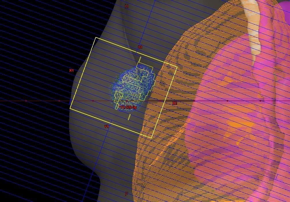
Radiation therapy for breast cancer
 Radiation therapy at EMC is performed using the latest generation of TrueBeam Varian linear accelerators, which allow targeted irradiation of the tumor with minimal impact on surrounding tissues. Such equipment is installed only in several dozen of the world's leading cancer clinics. The specialists of the department, under the guidance of the chief radiotherapist of Moscow, Dr. Nidal Salim, conduct radiotherapy treatment according to modern international protocols.
For the first time in Russia, EMC has implementeda unique method of treating early–stage breast cancer is organ-preserving surgical treatment with simultaneous intraoperative radiation therapy using the Xoft system (USA). The revolutionary technology allows radiation therapy to be performed in one session right during surgery, which significantly reduces the duration of treatment and minimizes the side effects of radiation.
Systemic drug therapy for breast cancer
Adjuvant therapy is a systemic antitumor treatment that is performed after surgical treatment. The purpose of adjuvant therapy is to prevent the growth of cancer cells that could spread throughout the body. Adjuvant systemic therapy is performed in most women with stage 2 of the disease and in some patients with stage 1.
There are three main types of adjuvant systemic drug therapy for breast cancer:
-
Chemotherapy. The decision on the need for chemotherapy is made by an oncologist based on a variety of factors, including the stage of the disease, the degree of tumor differentiation, the presence of hormonal receptors, etc. The main drugs in the treatment of breast cancer are doxorubicin or adriamycin, cyclophosphamide or cyclophosphamide, and taxanes. The group of taxanes includes docetaxel and paclitaxel.
-
Hormonal (endocrine) therapy. It is performed in patients with estrogen receptor-positive tumors, regardless of the stage. Reduces the risk of cancer recurrence by 50%. There are two main types of hormone therapy that are used in adjuvant treatment: antiestrogens and aromatase inhibitors.
-
Anti-HER2 therapy. It is recommended for patients whose tumors express the HER2/neu receptor. Trastuzumab and pertuzumab are used as targeted drugs.
Oncologists and chemotherapists at EMC carry out all types of systemic drug therapy of any level of complexity, using the latest drugs and methods of treating oncological diseases. The drugs are administered through the central venous access port in comfortable rooms. At the end of the chemotherapy or targeted therapy session, the patient can go home. At the request of the patient, a special cooling hose is used during treatment, which allows you to preserve hair during chemotherapy.
Treatment of stage 3 breast cancer (locally advanced cancer and inflammatory cancer)
 Stage 3 cancer is usually treated with a combination of chemotherapy, surgery, and radiation therapy. Hormone therapy and targeted therapy with trastuzumab (herceptin) may also be used. In most cases, systemic drug treatment is performed before surgery, which is called neoadjuvant therapy. In locally advanced cancer, mastectomy is more often performed than organ-preserving treatment, and almost all patients receive radiation therapy after surgery.
Stage 4 breast cancer treatment
In recent years, significant progress has been made in prolonging the lives of patients with metastatic breast cancer, while maintaining quality of life during treatment. Treatment methods are used that reduce the severity of symptoms and at the same time have minimal side effects on the body. As a rule, treatment tactics involve the reasonable use of systemic therapy. Surgery and radiation therapy can also be used for local treatment, for example, bone metastases, brain, spinal cord and skin metastases.
The prognosis for breast cancer
The five-year survival rate for breast cancer is:
-
at stage 1 – 95%
-
at stage 2A – 85%
-
at stage 2B - 70%
-
at stage 3A – 52%
-
at stage 3B – 47%
-
at stage 4- 18%
Most breast cancer recurrences occur in the first five years after diagnosis, especially in hormone receptor-negative tumors.
After breast cancer treatment
The therapy is followed by a period of follow-up medical supervision. At this time, the patient regularly visits the doctor, the regularity of visits is determined by the doctor for consultation. In addition, it is necessary to undergo an annual mammogram so that in case of a recurrence of the neoplasm, it can be diagnosed and treatment can begin at an earlier stage.
Make an appointment for a consultation and we will contact you for more details
Why the EMC
The first and only clinic in Russia, created in the image of the world's leading clinics


.webp)


Make an appointment for a consultation
Specify your contacts and we will contact you to clarify the details

Subscribe to the newsletter
Find out before others about the special offers
and new products of the EMC
and new products of the EMC