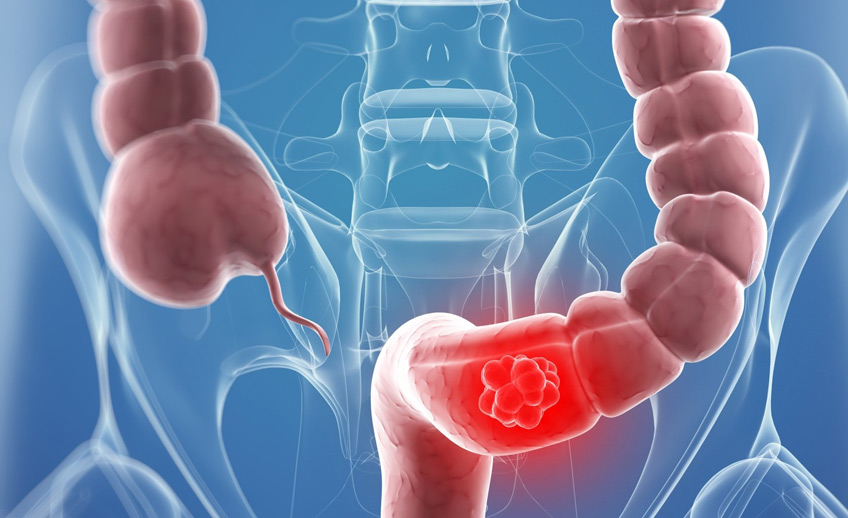
Colon cancer

The Institute of Oncology provides a full cycle of colon cancer treatment in accordance with international standards. Our patients receive all the necessary care in one clinic: from diagnosis to surgical treatment, radiation and drug therapy.
Colon cancer is one of the most common malignant diseases.In Russia, colon cancer ranks fourth in the structure of cancer incidence.The increase in morbidity
Colon cancer in most cases develops from
Colon cancer is more common in men. Age is considered a major risk factor for so-called sporadic (non-hereditary) colon cancer. In almost 70% of cases, colon cancer is diagnosed in patients&over 65 years old. Meanwhile, the disease practically does not occur before the age of 45.
Some intestinal diseases, in particular, Crohn's disease and ulcerative colitis;
Taking nonsteroidal anti-inflammatory drugs;
Smoking;
Hereditary predisposition.
It has been proven that diabetes mellitus, metabolic syndrome, abdominal enlargement, hypertension, hyperglycinemia, and low levels of high-density lipoproteins predispose to the development of colorectal cancer in men, but no such dependence has been found in women.Hereditary syndromes (familial adenomatous polyposis and hereditary non-polypous cancer syndrome) underlie 5-10% of all colon cancers.There is also a significantly increased risk of recurrence of a malignant intestinal tumor in those who have had colorectal cancer at a young age.
The purpose of screening is to detect precancerous diseases in a healthy population, as well as to detect malignant tumors at early stages.
Since age is the main risk factor, screening should be carried out for all men and women from 50 to 74 years of age. There are several screening techniques: stool analysis for hidden blood, fecal immunochemical examination, and endoscopy (fibrectoromanoscopy, colonoscopy). Analysis to detect feces for latent blood is recommended to be performed at intervals of 1-2 years, colonoscopy — at intervals of 7-10 years.
According to the results of three large-scale studies, a 25% reduction in mortality was demonstrated among those who took a stool test for hidden blood at least once. This served as the basis for the Committee on Cancer Prevention of the European Union to recommend a stool test for hidden blood as a screening, in case of a positive result— to perform an endoscopic examination.
The first symptoms of bowel cancer are easy to miss. Colon cancer in the initial stages is characterized by the absence of symptoms or its scarcity or non-specificity. As a rule, the clinical picture of the disease appears when the tumor reaches a significant size and it grows into neighboring organs. The patient may be concerned about changes in stool (constipation, diarrhea), general abdominal discomfort, unexplained weight loss, increased fatigue, anemia.
Advantages colonoscopies:
determining the exact location of the tumor,
specifying the volume of damage
biopsy sampling for histological examination
detection of synchronous precancerous or cancerous lesions
deletion polyps during the study
In
In cases where the right colon cannot be examined for anatomical reasons, as well as in patients with multiple adenomas, irrigoscopy is used in addition to colonoscopy.
Early diagnosis of colorectal cancer in EMC
When planning laparoscopic surgery, as well as to identify secondary tumors and adenomas in patients with stenosing tumors of the distal colon that prevent invasive examination, EMC uses additional techniques such as virtual colonoscopy is a CT scan of the colon.
To confirm the oncological diagnosis, a morphological examination of the biopsy material is necessary. At EMC, the study is performed by highly qualified histologists.The EMC Histological Laboratory cooperates with the American Society of Telepathology and leading cancer clinics in Europe and the USA. If necessary, or at the request of the patient, a second opinion from a foreign specialist can be obtained.
The standard assessment of the histological conclusion of the surgical material includes:
morphological description —determination of tumor location and size
presence or absence of macroscopic tumor perforation
histological types
the degree of malignancy
determination of the level of tumor growth into the intestinal wall and neighboring organs
the distance from the tumor to the resected edges
the presence or absence of a native tumor vascular or perineural invasion
the number of lymph nodes removed and their possible damage by cancer cells.
According to international recommendations, PET/CT scans are performed in patients with diagnosed colon cancer to detect distant metastases and determine further treatment tactics in EMC.
Staging of malignant neoplasms is carried out in accordance with the TNM classification. Stage determination is necessary to select the optimal treatment and select patients with single liver or lung metastases for surgical treatment.
Stage 1 — involvement of the submucosa and muscle layer. Absence of metastatic lesions of regional lymph nodes, absence of distant metastases.
Stage 2 is divided into sub-stages IIA and IIB.
Stage IIA —involvement of subseroses, nonperitonized areas of the colon/rectum
Stage IIB is the involvement of the visceral peritoneum or other adjacent organs and structures.
The second stage is characterized by the absence of metastatic lesions of regional lymph nodes and the absence of distant metastases.
Stage 3 is divided into three sub-stages:
IIIA —Involvement of the submucosa and/or the muscular layer.Metastatic lesion of three or fewer regional lymph nodes.
IIIB—Involvement of subseroses, non-peritonized areas of the colon/rectum, and/or involvement of the visceral peritoneum or other adjacent organs and structures. Metastatic lesion of three or fewer regional lymph nodes.
IIIC is any of the above-mentioned volume of involvement by the primary tumor. Metastatic lesion of more than three regional lymph nodes.
The third stage is characterized by the absence of distant metastases.
Stage 4 — any volume of primary tumor and any number of metastatically affected regional lymph nodes. The presence of distant metastases.
The examination before surgery includes:
examination by a doctor
blood tests (general and biochemical) with assessment of kidney and liver function
determination of the level of the REA cancer marker
computed tomography of the thoracic and abdominal organs, pelvis
performing a colonoscopy
Staging is also performed intraoperatively. The operation evaluates the condition of the liver, regional lymph nodes (at least 12 and 14 lymph nodes must be examined for adequate staging), and tumor growth into the intestinal walls and surrounding structures.
The determination of prognosis factors is necessary in order to form a further treatment plan, if necessary, to prescribe adjuvant chemotherapy after surgical treatment in order to reduce the risk of recurrence of the disease.
The prognosis for colon cancer directly depends on the depth of tumor invasion and involvement of regional lymph nodes. Also important prognostic factors are the degree of differentiation of tumor tissue, vascular or pleural invasion, and involvement of resection edges in the tumor process. Adverse signs include intestinal obstruction, tumor perforation, and increased levels of cancer markers CEA and CA 19.9.
The five-year survival rate after surgical removal of the tumor is as follows:
for the first stage – 85-95%
for the second stage – 60-80%
for stage 3 – 30-60%
Patients with isolated liver and lung metastases may undergo surgical treatment.
The five-year survival rate reaches 40% percent in patients after partial hepatectomy for single liver metastases.
Limited metastases in the lungs are less common than in the liver. Nevertheless, in carefully selected patients, after removal of lung metastases, the five-year survival rate ranges from 35 to 45%.
For stage 0 colon cancer, treatment involves local excision or polypectomy (endoscopically) or segmental resection.
At stages 1 and 2, low-risk patients undergo extensive resection with anastomosis.
The treatment of high-risk stage 2 and stage 3 also consists in wide resection with anastomosis followed by chemotherapy.
The volume of resection depends on the location of the primary tumor, the specifics of blood supply and distribution of lymph nodes, but in any case it is at least 5 centimeters from both sides of the tumor.
There are laparoscopic and laparotomic methods of surgical treatment. Recently, there have been many reports of colon cancer surgeries performed on the Da Vinci robot.
The main types of operations for colon cancer:
resection of the ileocecal angle, in which part of the small intestine and cecum are removed with the application of primary anastomosis (junction of the small and large intestine);
right-sided hemicolectomy, when the terminal small intestine and ascending colon with hepatic bend are removed;
resection of the transverse colon with adjacent lymph nodes;
left-sided hemicolectomy, when the splenic fold of the colon and the descending part of the colon are removed;
anterior rectal resection, when the sigmoid colon and the upper ampullary rectum are removed;
low anterior rectal resection - the upper and middle ampullary sections of the rectum are removed with resection of the sigmoid colon.
Most often, in a planned situation, primary anastomoses are formed — the two ends of the resected intestinal fragments are stitched together. If the operation is of an emergency nature, an additional removal of the stoma to the anterior abdominal wall is often performed — a colostomy or ileostomy.
Reconstructive surgery in patients with ileo- and colostomy
In some cases, when the patient is undergoing preoperative neoadjuvant chemotherapy or radiation therapy, and there are signs of partial intestinal obstruction, intracellular stents are installed using colonoscopy to preserve excretory function. After neoadjuvant treatment – radiation therapy, chemotherapy, or a combination of these two methods — the possibility of radical surgery is being considered.
In the case of the initial spread of the disease (the presence of distant metastases), surgery is performed to remove the primary tumor so that during adjuvant treatment (chemotherapy or radiation therapy or a combination of these methods), tumor tissue does not disintegrate or intestinal obstruction develops.
The EMC Surgical Clinic has extensive capabilities for early diagnosis and surgical treatment of benign and malignant diseases of the rectum, colon, and anus. Surgical consultations and treatment are carried out around the clock seven days a week.
Operations are performed by oncologist, coloproctologist, PhD Yuldashev Anvar Gafurovich.
The patient is admitted to EMC as planned on the day of the surgical intervention itself, except in cases where preoperative preparation in a hospital setting is necessary.
During the standard course of surgery, the postoperative period lasts from 3 to 7 days, depending on the patient's somatic condition (presence of concomitant diseases).
After the patient is discharged, bandages are performed, if necessary, both in the clinic and at home.
Radiation therapy (radiotherapy) is used in certain locations of the tumor process (rectum) or when the tumor spreads to neighboring structures and tissues. Radiotherapy is performed before or after surgery, often simultaneously with chemotherapy.
The EMC Radiation Therapy Center has the latest generation of linear accelerators that allow radiation therapy to be performed with extremely high accuracy and minimal impact on surrounding tissues. The department employs experienced specialists under the guidance of the chief radiotherapist of Moscow, Dr. Nidal Salim.
All patients with malignant tumors who have completed treatment require dynamic monitoring. Patients who have been treated for colorectal cancer undergo follow—up examinations every 4 months: CT of the abdominal cavity with intravenous contrast, low-dose CT of the chest, and colonoscopy every 8 months.
The most significant predisposing factors
Screening
Symptoms of colon cancer
Colon cancer diagnosis
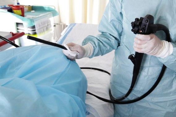 Colonoscopy, an endoscopic examination that allows visualization of all parts of the colon, is the most accurate and informative diagnostic method.
Determining the stage of colon cancer
Prognosis for colon cancer
Colon cancer treatment
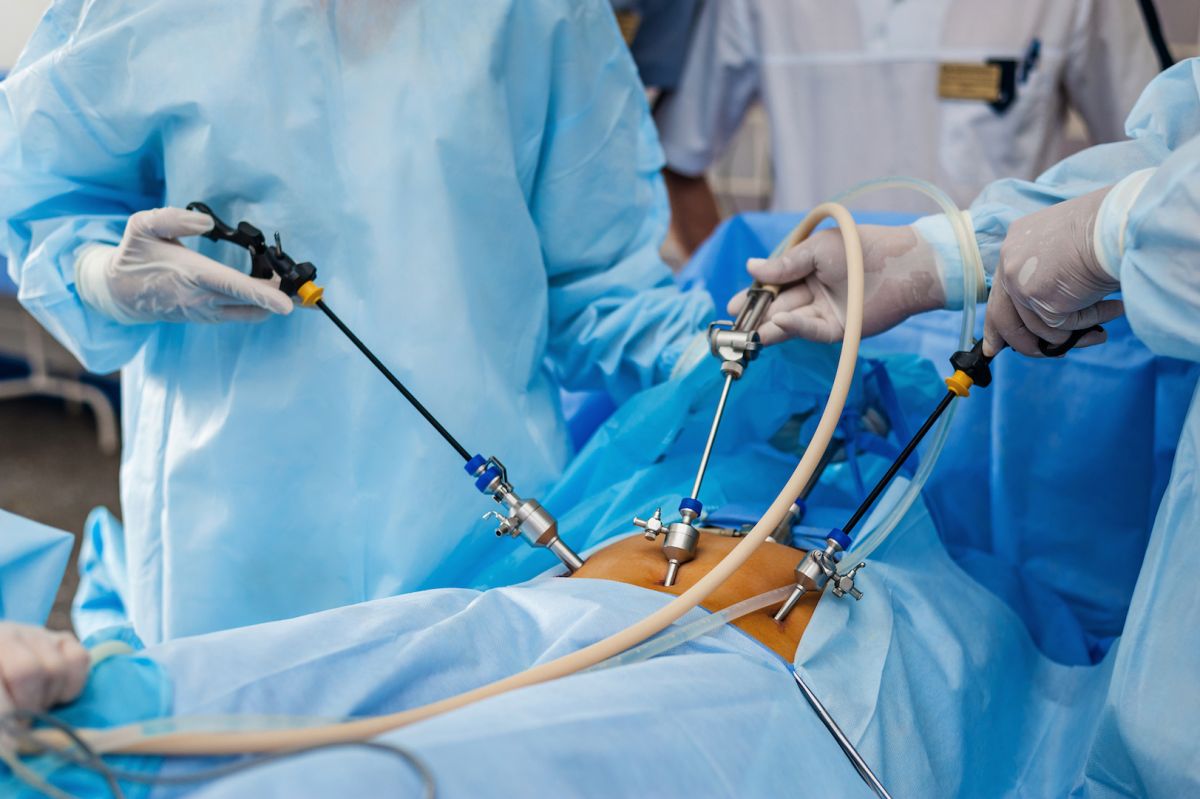 In the early stages (low-risk stages 0, 1, 2), surgical excision is the treatment method for colon cancer. The objective of surgical treatment is a wide excision of the colon segment along with the lymphatic collector.
Surgical treatment of colon cancer
Stenting for colon cancer
Features of surgical treatment in EMC conditions
Radiation therapy
Why the EMC
The first and only clinic in Russia, created in the image of the world's leading clinics


.webp)


Make an appointment for a consultation
Specify your contacts and we will contact you to clarify the details

and new products of the EMC