Cervical cancer is a malignant neoplasm that occurs in the lower part of the uterus and often metastasizes to other organs. For more than ten years, the European Medical Center has developed the highest expertise in organ-preserving operations, which are prescribed in the absence of contraindications and help patients maintain fertility. An integrated approach to diagnosis and treatment often allows patients to receive a positive prognosis not only in the early stages. If we talk about the general statistics of patients with cervical cancer in the USA and Europe, then 80-85% of remission occurs at the first stage, 70-75% at the second, 45-50% at the third and 5-7% at the fourth. When choosing where to treat this disease, you should pay attention to the clinic's statistics. The percentage of achieving long-term remission in patients with cervical cancer at the European Medical Center is higher than the general statistics due to more progressive and selective approaches to the treatment of different stages of the disease.
Types of cervical cancer
There are two types of cervical cancer, depending on the type of cells that are affected by HPV infection. Squamous cell carcinoma is most common. In this case, the tumor develops from the superficial epithelium that covers the cervix from the outside. The second type of cancer is glandular, or adenocarcinoma: this type occurs in only 15% of patients and arises from the inner layer of the cervical canal.
Causes of cancer
Most often, women aged 41-43 suffer from cervical cancer. The main and perhaps the only proven cause of cervical cancer is high–risk HPV (human papillomavirus) infection. In addition, there are additional risk factors, such as smoking.
Women who start having sex early and have many partners have a two- to three-fold increased risk of developing cervical cancer compared to others. Previously, it was believed that cervical cancer is caused by background diseases (erosion, leukoplakia, and others). But now it is known for sure that only the presence of HPV infection significantly increases the risk of developing the disease. Without HPV, infection with cervical cancer is very unlikely, even in the presence of the above diseases
.
As with any cancer, in the first stage, the tumor is located within the organ in which it originated, in this case, in the cervix. The first stage is divided into two sub-stages. Microscopic (1A) is established when the diagnosis of "cervical cancer" is detected only by the results of a biopsy. The histologist indicates how deep the cancer has spread (in millimeters), and this determines the specific stage. If the changes are visible during examination in the gynecological chair, this is already a sub-stage 1B. In this case, a biopsy is performed in order to histologically determine the type
of tumor.
In the second stage of cervical cancer, the neoplasm spreads to neighboring structures: the walls of the vagina or more deeply located lateral ligaments of the cervix – parametria. A doctor can detect a tumor during examination in a gynecological chair, determine its prevalence, and magnetic resonance imaging of the pelvis with contrast (MRI) helps to clarify the details of the lesion. Positron emission computed tomography (PET-CT) is also crucial in order to understand whether the tumor has spread beyond the pelvis – into the abdominal cavity, lungs, distant lymph nodes and other organs. This helps to correctly identify the fields for the upcoming radiation therapy and "capture" all tumor formations in the irradiation zone.
In the third stage, the tumor further affects neighboring organs and can cause blockage of the ureter, and in the course of progression, kidney failure. In this form of the disease, the tumor can compress large vessels and nerves in the pelvic area, causing pain and swelling in the legs. The lesion of the pelvic lymph nodes is another characteristic manifestation of this stage.
The fourth stage may be local, when the tumor does not grow sideways to the vessels and ureter, but forward towards the bladder or backward towards the intestine. If the neoplasm has grown into the bladder or intestines, this is stage 4A. If it has spread to the lungs, liver, bones, it is 4B. The probability of tumor recurrence after the end of treatment depends on the stage at which the cancer was detected. The higher the stage, the more likely it is to get sick again.
Advantages of cervical cancer treatment in EMC

An international and interdisciplinary team of world-renowned experts and the opportunity to get a second opinion from foreign doctors

Effective targeted non-surgical treatment

Chemoradiotherapy using the precision innovative
RapidArc/IGRT technique

The possibility of performing organ-preserving operations in the early stages of cervical cancer, follow-up, planning and management of pregnancy by obstetricians and gynecologists and other experts
Surgery for cervical cancer. Organ-preserving treatment
At the first stage of the disease, in the case when the tumor of the cervix is no larger than two centimeters, it is often possible to perform one of two options for organ–preserving operations - knife conization or radical trachylectomy. Many women do not have children at the time of diagnosis, and this type of treatment allows them to become pregnant in the future. At the earliest, microinvasive stage, when the tumor has only superficially grown to three millimeters into the cervix, it is possible to safely remove part of the organ through the vagina. This type of operation is called knife conization. If subsequent histology data confirms that the cancer cells have been completely removed, then such an intervention will be sufficient, and after six months the patient will be able to plan a pregnancy.
If the tumor is not at an early stage, but is less than two centimeters, the patient is offered a more complex organ–preserving operation - a radical trachylectomy. During it, the entire cervix with the surrounding vaginal tissues, parametria and lymph nodes is removed. In this case, the uterus with appendages can be preserved, which means that the patient will retain her reproductive function. Approximately 75% of pregnancies after the procedure last until the third trimester, and the baby is born already quite strong. Such organ-preserving operations have the same oncological prognosis as radical
ones.
If the tumor is large and/or there is no goal to preserve organs, radical treatment is necessary: surgery, chemoradiotherapy or radiosurgery. Since most patients with cervical cancer are young women, it is important to maintain ovarian function. We offer our patients special laparoscopic surgeries in which the ovaries are moved from the pelvis to the abdominal cavity, so they do not fall into the zone of future radiation.
If our patients want to become mothers in the future, we do everything possible to preserve their reproductive potential, and we offer either organ-preserving operations or freezing of oocytes and preservation of ovarian tissue. If pregnancy is not planned, radical surgical interventions can be performed – removal of the cervix along with the uterus, the upper part of the vagina, the parameters and lymph nodes. Minimally invasive surgery (laparoscopic or robotic surgery), at first glance, seems to be the best choice for treating cervical cancer. But a recent competent and multicenter international study has shown that this type of treatment significantly worsens the prognosis and does not exclude the occurrence of relapses. Therefore, EMC specialists recommend open surgeries: for more than ten years, Vladimir Nosov, PhD, Head of the Clinic of Obstetrics, Gynecology and Oncogynecology, oncogynecologist, and the only full member of the American Society of Gynecological Oncologists in Russia, has been working on this issue. He works in a team with highly qualified colleagues – oncologists, reproductive specialists, radiation diagnosticians, embryologists, psychologists.
Advantages of the EMC operation:
- Organ-preserving operations
-
Preservation of reproductive potential, the possibility of motherhood
-
The operations are performed by Vladimir Nosov, PhD, Head of the Clinic of Obstetrics, Gynecology and Oncogynecology, oncogynecologist surgeon, the only full member of the American Society of Gynecological Oncologists in Russia
Non-surgical methods in the treatment of cervical cancer
The second, third, and fourth stages are considered inoperable, but this does not mean that permanent remission cannot be achieved in such cases. Patients of the second (inoperable stage) without surgery, they go into remission in 75-80% of cases, and even more often within the walls of the EMC. Inoperable only means that surgery at these stages is not the optimal treatment option, unlike chemoradiotherapy. According to classical international studies, weekly administration of small doses of chemotherapy in parallel with ongoing radiation therapy leads to an improvement in the cure rate by almost 10% compared with standard radiation therapy.
Radiation therapy
Radiation therapy is the main method of treating cervical cancer of the second and third stages, the course of treatment lasts 7-8 weeks. Detailed prices for a course of radiation therapy for oncology at the EMC clinic. With this treatment, daily sessions are held from Monday to Friday, for 15-20 minutes. There is a standard approach when remote radiation therapy is performed for five weeks at the first stage. In this case, the irradiation occurs without contact with the patient's body. As is correct, on Mondays, in addition to radiation therapy, a drug with chemotherapy is injected through a venous catheter for an hour.
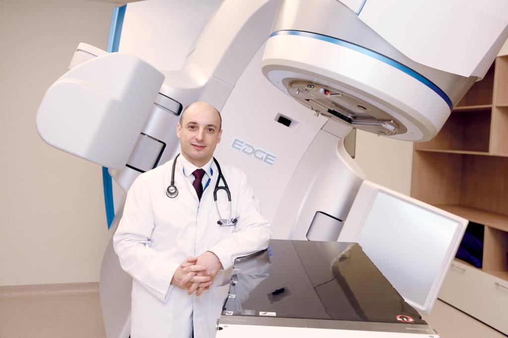
Remote radiation therapy in its standard form is not always effective. During the procedure, both tumor-affected and healthy tissues are irradiated: as a result, the tumor is not completely destroyed, because the permissible radiation level is not high enough. Healthy tissues receive a radiation burn. Five weeks later, the patient undergoes brachytherapy sessions — contact radiation therapy. During the procedure, a special device is inserted into the vagina, which locally irradiates the surrounding tissues, including healthy ones, for 40-50 minutes. As a rule, treatment involves 4-6 sessions of brachytherapy with an interval of several days — this is the so-called boost. The procedure is quite painful, so it is usually performed under sedation.
Even 15 years ago, oncologists all over the world were very concerned about the insufficiently satisfactory results of such non-selective treatment with colossal toxicity. An alternative technique has emerged in which radiation therapy is delivered only to tumor tissues, without affecting healthy ones. This method involves long and complex radiation planning by a team of doctors and radiophysicists for many hours. The radiation unit is programmed with a completely unique system for each patient, which allows it to affect only the tumor, giving doses significantly higher than the standard ones. Moreover, boost (additional irradiation of individual areas) is integrated into the remote therapy program, and the use of vaginal implants for brachytherapy is not required.
At EMC, we have been offering our patients just such a highly accurate and precision treatment method with minimal or no side effects for many years – Volumetric Modulated Arc Therapy Intensity-Modulated Radiation Therapy (VMAT/RapidArcIMRT). The very first patients we treated in the late stages of cancer using this radiation technique are still in remission after years of follow-up and maintain an excellent quality of life. Previously, it was difficult to imagine such a thing, but today it is a reality for EMC patients. Few patients receive such an individual treatment plan outside the EMC, and most continue to fight cervical cancer according to the general scheme. But treatment depends not only on a high level of technological equipment, but also on a personal approach to each patient and on the experience of radiotherapists.
Advantages of radiation therapy in EMC:
-
A special method of radiation therapy Volumetric Modulated Arc Therapy (VMAT/RapidArc), Adaptive non-brachytherapy approach – healthy tissues do not suffer
-
A team of world-class doctors and radiophysicists
Chemotherapy
In combination with radiation therapy, small, so-called half-doses are used for chemotherapy. If the disease is detected at the fourth stage, treatment often begins with chemotherapy, sometimes targeted drugs are also involved. This makes it possible to reduce the size of the tumor: when it gets smaller, radiation therapy, including stereotactic radiosurgery (SBRT), is added to the treatment.
Advantages of chemotherapy in EMC
At the EMC, we use certified foreign drugs for chemotherapy. There are young women with reproductive plans who still have a tumor in the first stage, but it is already larger than two centimeters. In this case, during treatment, we can first safely reduce the tumor to a size of less than two centimeters with the help of preparatory chemotherapy, and then perform surgery with the preservation of organs. With this treatment, we select drugs that will have a minimal effect on the ovaries and will not reduce her reproductive potential.
Targeted therapy
In cervical cancer, targeted medications are used in combination with chemotherapy and immunotherapy. This method allows you to purposefully destroy cancer cells without significant harm to healthy ones.
Advantages of targeted therapy in EMC
The treatment of cervical cancer with targeted therapy is available to EMC patients. We have the opportunity to use immunotherapy drugs that target specific molecular or immunological breakdowns: we detect them in tumor tissue. Then we make up her molecular genetic portrait and select a special drug that will affect only the tumor cells.
Cancer symptoms
At an early stage, the malignant tumor is located in the cervix and does not go beyond its borders. As a rule, a woman develops abnormal discharge – bloody or mucous, sometimes with an unpleasant odor, and there may also be a malfunction of the menstrual cycle. At the same time, painful sensations are often absent, and suspicions of the disease appear only during an examination by a gynecologist. In the later stages, a woman's leg often swells on one side, she experiences shooting pains that radiate to the buttocks. Sometimes the outflow of urine is blocked, and therefore the back begins to hurt.
Diagnosis of cervical cancer
The diagnosis consists of several important stages that allow you to accurately diagnose and make a competent treatment based on it. This is a cytological smear, a biopsy of the cervical tumor or the cervix itself if early cancer is suspected, followed by an MRI of the pelvis with contrast and PET-CT.
Screening
A cytological smear or PAP test is a screening examination. It is given to all healthy women to identify potentially pathological cells. If they are found, women are invited for a more detailed diagnosis (they examine the cervix under a microscope and take the material for a biopsy) in order to confirm or deny the diagnosis of a precancerous condition or early cervical cancer. It is recommended to take a cytological smear from the cervix for the first time at the age of 21, and the first high–risk HPV test at the age of 30.
There is a phenomenon of HPV self-elimination: in 80% of cases, when the virus enters the female body before the age of 30, it leaves it on its own. After the age of 30, this rarely happens, so it is important to monitor the presence of HPV. The recommended frequency of examinations is once every 1-3 years, so as not to miss early changes, but we advise our patients to be monitored annually.
PET-CT
If the results of the control MRI examination do not allow us to judge with certainty the absence or presence of glioblastoma progression, the patient is prescribed a PET-CT scan of the brain with an amino acid-based radiopharmaceutical.
The EMC Clinic is the leader in PET technology in Moscow and uses 18F-fluoroethylthyrosine (18F-FET) for this purpose, which has proven its advantage in numerous studies, allowing it to detect tumor progression in complex diagnostic cases, allowing timely and adequate response to the current situation, up to the need for repeated neurosurgical intervention.
The price of cervical cancer treatment in Moscow at the EMC clinic is calculated individually after consulting an oncologist.